Polymeric Separations
This post outlines two sets of polymer membrane separations using 100kD PES and 30kD cellulose in diffusion mode. The 100kD PES experiment is a repeat of the experiment found here; in the first trial of this work, there was no filtrate remaining on the backside. This time there was filtrate remaining that I could test for protein. You can find results for 100kD cellulose and 50kD cellulose on the linked pages.
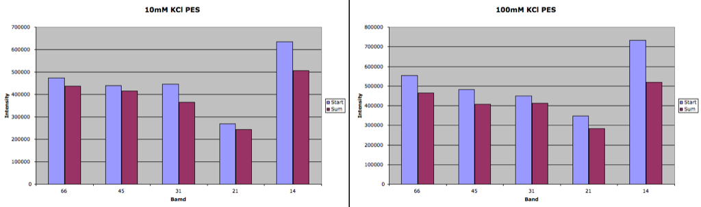
100kD PES:
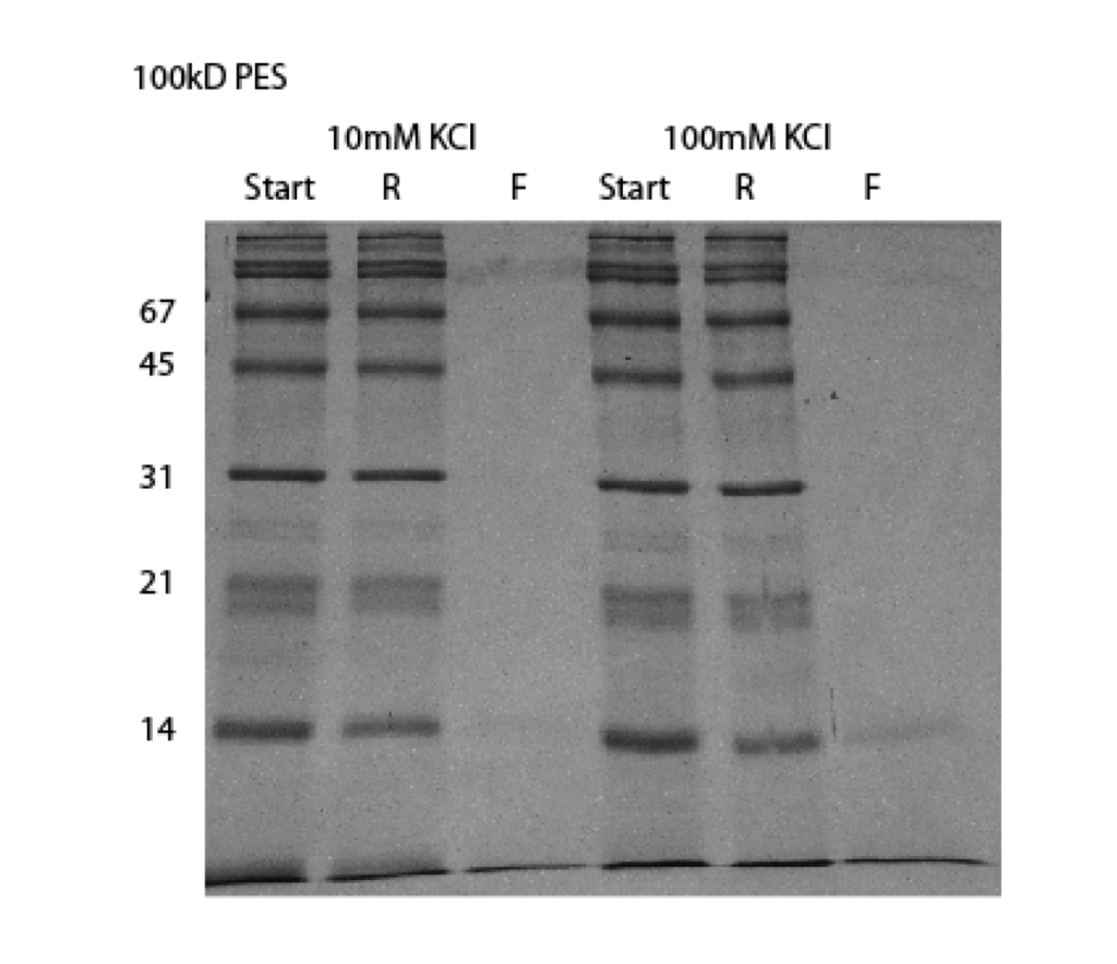
There were no visible proteins in the filtrate from this separation. These membranes are not made for diffusion, and it obvious that they do not function well in this mode. This may be due to the more hydrophobic nature of PES. 100kD cellulose was capable of separating the protein in diffusion while 100kD PES is unable.
If we use our previous method to test for loss, the following graphs show there is little difference between the starting solution and the sum of the retentate and filtrate. The loss is probably within the noise. I’m not sure why there was so much more loss in the previous PES separations. There was really no difference between the 10mM and 100mM KCl separations in this work.
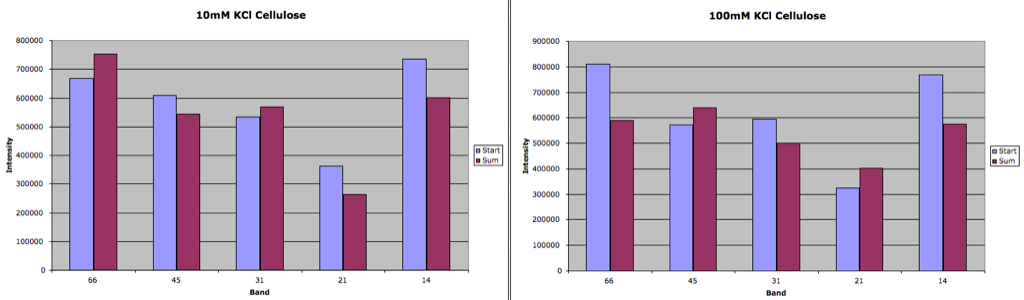
30kD Cellulose

It seems that there’s a couple of bands in the 10mM separation that are not visible in the 100mM separation, however it’s possible that this is a leaking or dud membrane. Again, these membranes are not made to be used in diffusion. The losses appear to be mostly minimal.
So there may be a unique opportunity for pnc-Si membranes that can fractionate small protein mixtures.
Can you generate a table with then native size, gel MW, and name of each protein in the ladder?