BSA diffusion across SC267
Last week I made a post showing detection of detergent diffusion across membrane SC267. In the diffusion set up I had placed 6uL of a 50/50 volume ratio solution of 1.0% DDM and 1mg/mL BSA with 60uL of DI-water on the membranes backside. I allowed the setup to sit for two days and then used a colorimetric assay to determine the amount of detergent that diffused across. The problem with last weeks experiment was that I could not determine whether BSA had also diffused across the membrane.
I ran this experiment again except this time I use four SC267 samples; two with 6uL of 50/50 volume ratio solution on top, one with 6uL of only 0.5% DDM on top, and the last with 6uL of only 0.5mg/mL of BSA on top. I again allowed the setup to sit over night before collecting the filtrate and running the appropriate assays.
To detect for the presence of DDM diffusion across the SC267 samples who started with the 50/50 volume ratio solution and the 0.5% DDM solution I used the phenol/sulfuric acid colorimetric assay. As shown below the assay detected the presence of detergent in the filtrate in two of the three tested samples.
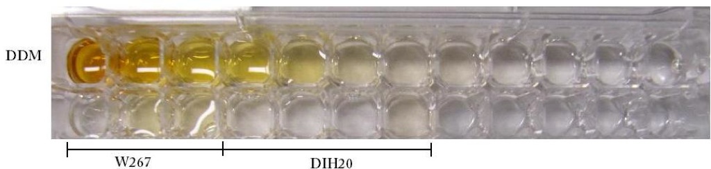
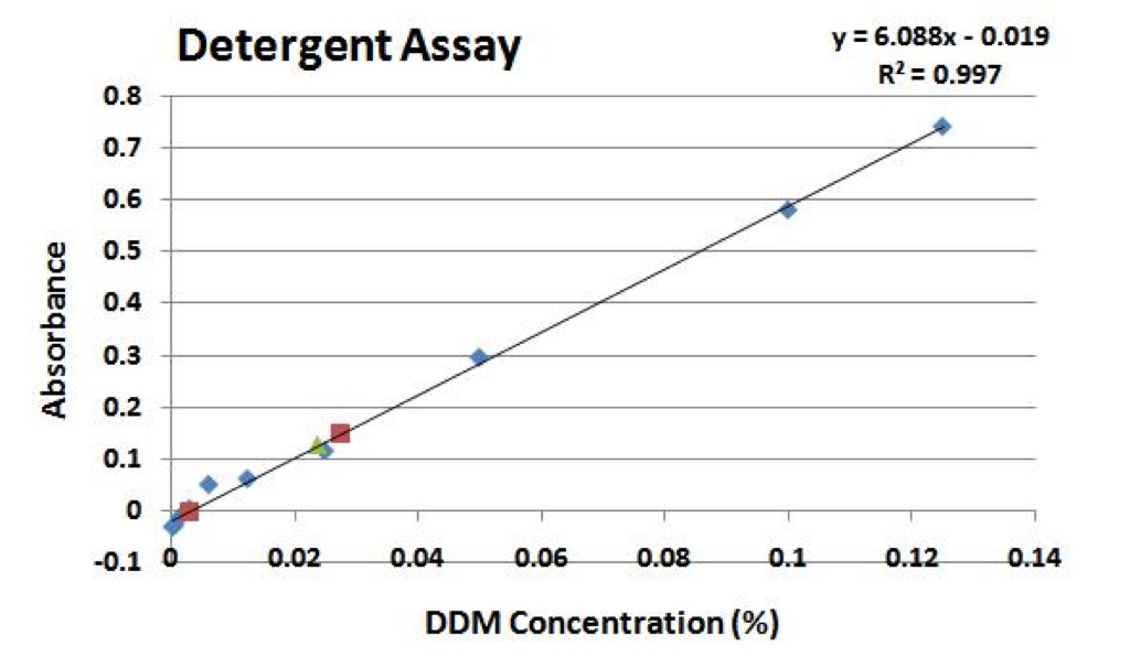
The red squares on the plot above indicate the DDM concentrations calculated for the samples that started with 50/50 DDM/BSA on the top of the membrane. These concentrations were determined to be 0.003% and 0.027% respectively. The green triangle on the plot above indicates the DDM concentration calculated for the sample that started with only 0.5% DDM on the top of the membrane; its concentration was calculated to be 0.024%. It should be noted that the original detergent concentration on the top side of the membrane in all three diffusion sample setups was 0.5%.
To detect for the diffusion of BSA across the final SC267 sample a bradford assay was used. The image below shows the results of this assay.
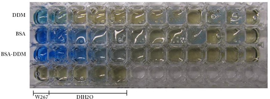

The above plot graphs the linear detection region for the bradford assay on BSA. The red square indicates the calculated BSA concentration in the samples filtrate (0.038mg/mL). It should be noted that the initial BSA concentration in this diffusion setup was 0.5mg/mL.
Doing the math, the conversion from 6uL on the topside of the membrane to 60uL on the bottom should give a 1:10 dilution at equilibrium. For the DDM we see a dilution from 0.5% to ~0.025% or a 20-fold dilution. On the other hand, for the BSA we see a dilution from 0.5mg/mL to 0.038mg/mL or a 13-fold dilution. From these results it does not appear that BSA diffusion is being hindered by the charge on the pnc-Si membrane.
I would appreciate any thoughts or input on the matter, but I think my next step is to try to repeat the experiment possibly using oxidized samples.