Vacuoles via SEM
I had some extra samples from last week and had been wanting to try SEM on cells, so I went for it this week. Specifically, I was hoping to get images of vacuoles in cells over free-standing pnc-Si. I followed a rather complicated protocol full of dangerous chemicals to fix the cells and then dehydrate them into an ethanol series and then an HMDS series. I coated the cells with 6nm gold and then went to the SEM. Right after I got on the SEM, the chilled water supply was taken down to fix a leak, so I got kicked off. I was able to get 1 scan:
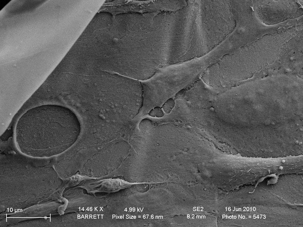
The bright spot on the upper left is a folded over, free-standing membrane, and I believe the crease running almost vertical around the center of the scan is the free-standing to supported interface. It looks like there are cells of various morphologies on the membrane, with 1 enormous hole and several smaller holes present. I believe these are vacuoles. This is not a high enough magnification to see the pores underneath the vacuole, however. Since I was kicked off and most of my samples did not survive the fixing/drying protocol, this was the only piece of data I got from today’s SEM. However, it looks like my protocol works – I will try again with pseudo-transwells so the pnc-Si is easier to manipulate.