Cadherins in epithelial cells
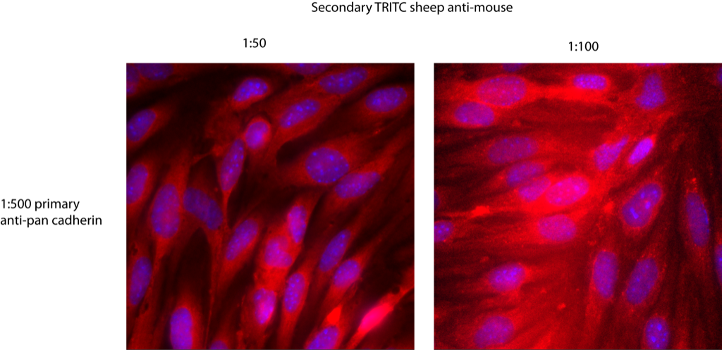
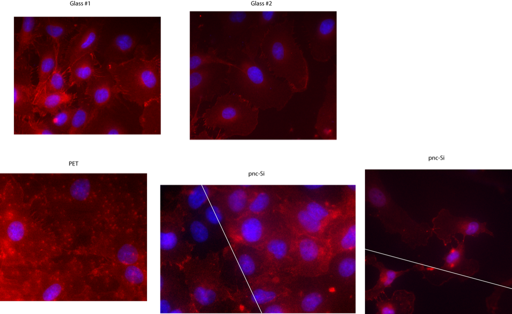
Those with a great memory will remember that I’ve been struggling (1, 2, 3) to identify adherens and tight junction proteins (i.e., cadherins and ZO-1) in bEnd3 endothelial cells. Since I’ve recently moved over to investigate air-blood barrier co-culture models, I decided to see if my epithelial cells (RLE-6TN) express cadherins (they should since these are also barrier cells). For this experiment, I grew RLE-6TN on glass, pnc-Si transwells and PET transwells for 1 day only in order to help prevent cell multi-layer formation. I then did cadherin immunofluorescence to stain cadherins red and counterstained with Hoechst 33342 before imaging at 100X on the Zeiss. These cells show cadherin expression and distribution:
Cadherins are localized to the leading edges of cells; in neighboring cells, they are expressed at the cell-cell junctions. Importantly, the distribution of cadherins is similar on glass, PET and pnc-Si, which indicates that these cells are maturing normally on transwells. Additionally, cadherins can be seen in cells both on supported and free-standing pnc-Si (to the right of the white line is free-standing pnc-Si). Fluorescence background for pnc-Si and glass is negligible in all channels but significant for PET (so significant that it is difficult to identify cadherins on this sample). In fact, the puncta of red fluorescence in the PET image are the 400nm pores in this membrane (I confirmed the source of this autofluorescence in PET samples without cells). Therefore, cadherin expression is similar on pnc-Si and glass. Also, image quality on pnc-Si is much greater on pnc-Si than PET.
Updated on August 25th to include a new panel of RLE on pnc-Si (supported pnc-Si on bottom). Note the reduced background – I’d say identical to glass. This shows that pnc-Si background (and image quality) can match glass for high resolution immunofluorescence.
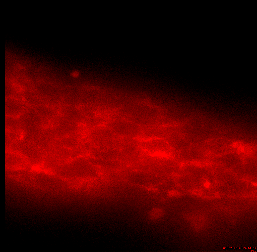
There is a brighter background fluorescence over the free-standing pnc-Si area, which I’ve noticed with many different fluorophores and on the Nikon, Zeiss and confocal scopes. I believe this is reflection of fluorescence but I’m not sure if it is a phenomenon attributable to nanometers thick pnc-Si or some weird reflections in the wells. Here is an image of cells on supported pnc-Si, which shows a little less background fluorescence:

In a different experiment, I stained RLE cells for cadherins when they were cultured on the well side of pnc-Si transwells. Unfortunately, these cells were ~ 4 days into the culture and so a huge multilayer was present and the fluroescence was blurry. However, you can see a more robust and defined cadherin expression around the periphery of individual cells (at cell-cell junctions):