Initial Testing of HUVECs w/ CMFDA
When the HUVECs were first brought up, the first test for me to perform was to see how they reacted to CMFDA and repeated fluorescent excitations. Based on the implications from previous research (summarized here), there is the possibility of harm or phototoxicity from the dye. Since HUVECs are more valuable and ‘fragile’ than either CHO or bEnd.3 (the previously used lines), knowing if CMFDA would have an impact is important if the dye is to be used for any sort of tracking or aid in other experimentation.
The results seem to imply that there is an effect on the HUVECs as well, though not as severe as is evident from the bEnd.3 pictures. There is still reduction of cell spreading and ‘happiness’, as well as apparent inhibition of proliferation. While it does seem that there are a number of cells still surviving after almost two days in the least favorable experimental conditions (100% intensity excitation given each timepoint), they do not appear to happily growing and there is definitely no signs of a confluent monolayer in that area. For any experiments where viewing cells repeatedly to follow angiogenesis, proliferation, etc, I would recommend either using phase microscopy or finding another dye with less apparent harm and/or variability.
This experiment was performed similarly to the final experiment on the previous blog post. It was also performed on a similar time table (46.5 hours) as was the other experiment (with bEnd.3; 48 hours). Five wells in a 24 well plate were seeded with HUVEC cells, three containing CMFDA and two simply being seeded as per standard protocol. Three wells served as controls. One well without CMFDA also had no exposure to any excitation (only basic ambient light and that needed for phase microscopy was used). The second well without CMFDA was exposed for ~5 seconds to excitation each time images were taken. The third control well had CMFDA, but received no fluorescent excitation.
The two wells which did have CMFDA and were being excited differed mainly in intensity of the light. One well experienced 100% relative intensity, while the second received only 17% (but had a longer exposure time).
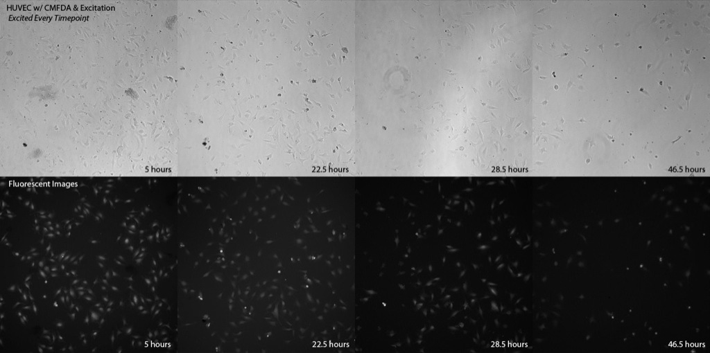
The images for each trial in phase and fluorescent (where applicable) are listed with times, and all were taken at 10x. Also, in the first images there was a fairly large amount of non-fluorescing debris, which was later determined to most likely be either cellular blebs from apoptosis, or potentially large aggregates of proteins (but it does not seem to have hampered cell growth and viability).
The normal control, with neither CMFDA nor excitation, grew as has been seen before with adherent cells. While not reaching full confluence in the two days observed, they did grow towards that end and did appear to be well-spread for the most part.

Similar to the previous experiment, it would appear that adding CMFDA without exciting it does nothing to the cells’ ability to grow and proliferate. This location did appear to start at a higher density than optimal to view proliferation, but it is apparent that the cells still spread out further and filled in whatever space there had been. In addition, even after the 46.5 hours only a small percentage of cells were rounding or showing signs of death (as is normal once confluence or near-confluence is reached in a given location).

When exciting the cells without CMFDA, there did not seem to be any hampering to proliferation caused by the light itself. Cells appeared viable and abundant, and followed the expected growth trends. The exception of the location that was excited all but the first timepoint (second row, below) could have been caused by the evident granules floating about, which are mostly absent from the other sets of images after the 5 hour mark. I am not entirely sure what they are, but it is unlikely that they are live contaminant since a) they are not ubiquitous in the well and b) even if consistently present at that location, do seem to decrease in concentration over time.

The first of the experimental results with CMFDA & excitation light seems to follow a similar trend as before with the bEnd.3 cells. While not as rapid or strong an influence against the cells, it appears that exciting the dye every timepoint still impeded cell growth and caused gradual roundedness of cells, implying death or at least negative conditions for the cells’ viability.
When not exciting on the first timepoint (waiting until the 22.5 hour mark), it seems the cells were able to grow and thrive more happily. While not proliferating as heavily as may have been expected (which may have been caused partially by the initial debris), they still grew and remained adherent and spread, marking a viable environment.

Exciting the cells at only the first and final timepoints seems to have had a more negative effect than merely skipping the first. Again, while not as harmful it seems as exciting every time, there still was reduction in spreading of cells and slightly reduced confluence.

The rest of the images were taken in a well where the duration of excitation was equivalent, but intensity greatly reduced (to 17%). Camera exposure time was boosted 4x to allow for better viewing of the fluorescence, with EM Gain left at 0. The first set of images, representing a location which was excited at every timepoint, seems to follow even more closely the trend shown in the bEnd.3 experiment. Cells which started in the area of interest began rounding and becoming unhappy, and didn’t proliferate well. Ultimately, cells from outside the area being imaged seemed to ‘invade’ into the space to use the empty space (see top-left portion of images). It would appear, however, that despite the negative response that the cells began to rebound, as shown by cells in the bottom of the image that began to spread out again after initially beginning to round up.

When choosing not to excite at the first timepoint, the cells seemed to proliferate and live well, growing to confluence within the two days and showing low percentage of rounding or death.

In the case of 17% intensity and taking pictures w/ excitation only at the first and last timepoints, it would appear the cells were mostly happy and thriving well. While not growing to full confluence, they also started at slightly lower density than the previous location, and still become ~90% confluent.

It seems that CMFDA does have potential to hinder cells, including CHO, bEnd.3, and the HUVECs which are now the main focus of most of the cell culture in the Gaborski Lab. Even if the effects do not fully kill or stop the cells from proliferating, it seems from this data that there is enough of an influence as to be too large a factor if some sort of angiogenic assay or other experiment were to be performed with the CMFDA. It does look to be that if one does not repeat locations at all and merely jumps to different places in a given well to image, that the cells are fine and will grow properly, but this would negate the ability to track cells, or do something like watch tube formation in a given spot with fluorescence.
I have emailed Invitrogen and some other researchers who have either noted issue with CMFDA or had success with other dyes. So far one individual has responded, stating that they use PKH26 dye to track their cells for up to 8 days, which considering our present research would be more than enough. This is also a generic dye, and would work with most (if not all) mammalian cell lines.
Regardless of what steps are taken forward (whether obtaining a new dye, employing use of something like an antioxidant or radical scavenger as Kevin suggested, or just not using fluorescence for cell culture), it seems that CMFDA is not a fully viable option for all of the research focuses in the lab.
I am assuming you have a shutter on your light source (if bulb-based) or pulsing your LED’s so the sample is only exposed while the image is being acquired?> If you have an EM-CCD camera (you say EM gain is 0 in these experiments) try shortening your exposure by using this useful feature.
There are a couple of other options red-shifted towards the more biocompatible end of the visible spectrum, for instance CMTPX, CMTMR, CMRA. Good general info here:
http://tools.invitrogen.com/content/sfs/manuals/mp02925.pdf
There is a shutter, however I did want to equalize exposure and so allowed a slightly longer than normal time (~5 seconds) for each sample to be exposed. The exposure time difference for the lower intensity was a setting to allow the camera to pick up more fluorescence, but the beam was on the same amount of time.
I had wanted to use no EM Gain to equalize across the board, but employing that (which is an option) could be helpful, yes. Thanks for the suggestions, and your comment on the other CMFDA post (I did use the lower intensity from that suggestion).
And thanks also for the push towards a more red-shifted stain. I had considered PKH26 (which I do believe is closer to the red end also), but will take a look at the possibility of using other dyes if we indeed want to move forward with fluorescing of the cells over extended periods of time.
Thanks again!