HUVEC Growth on Various Substrates
In evaluating the usefulness of our membranes for cell culture, it is important to understand if and how cell growth is affected by the surface. The desire is that they be consistent with standard culture substrates (i.e. Tissue Culture Plastic (TC)).
I performed two growth trials using HUVECs (Human Umbilical Vein Endothelial Cells), one focusing on the 3.0µm pore size, 300nm thick SiO2 membranes. The other looked at 0.4µm pore size, 300nm thick SiO2 membranes.
For the first trial (3.0µm), controls consisted of TC and glass. For the second (0.4µm), only TC was used as a control.
In each case, three types of coating were used. A thin Collagen coating (~0.05mg/mL), a thin Geltrex (basement membrane protein mixture) coating (~0.156mg/mL), or no coating at all were used.
By ‘thin coating’, I mean a dilution of the protein(s) which prevents gel formation, but still allows a deposit of protein on the surface for adhesion and proliferation benefit.
It seems that in both experiments, only the Geltrex coated membranes were able to sustain proper cell adhesion and growth. For 0.4µm, it appears that the higher pore density allowed for best cell growth. For 3.0µm, it seems to be that the low pore density provided a better condition for growth. It may be that the smaller pores hinder/affect growth less than the larger pores.
In all cases, the substrates were housed in a 24 well plate. For membranes, a single-well CytoVu with 1000µm gaskets were used (and placed directly in one of the 24 wells). For glass, a PDMS ‘inverted stencil’ was used in conjunction with a diamond-tipped pen to cut a circle out of an 18x18mm #1.5 glass coverslip which could fit in the well. A piece of PDMS was placed underneath to help stabilize and prevent undesired shifting and floating. Two of the ‘top’ gaskets used in a single-well CytoVu (open area of roughly ~3.75×3.75mm) were placed on the glass circle prior to seeding to normalize for growth area. For TC surfaces, the bottom of the 24 well was used (also restricted by placing two of the ‘top’ gaskets/well for use as the growth area). Basic setup is shown in this schematic (glass has the same setup as TC):

After allowing ~1 hour and 10 minutes after coating, cells are seeded at a density of 5*10^3 cells/cm^2.
Images were taken either once or twice per day, and after the first set of images (~4 hour 15 minutes after seeding), the wells are flooded with ~750µL of media (Medium 200 is used for these experiments). When refreshing the media (done roughly every other day), it is aspirated out and another 750µL is placed down. To avoid damage to the membranes, aspiration was done underneath the chip (might have affected cells?).
After all the data was collected, either Dave or myself counted the cells in the images and charts were compiled based on the data (the final two data points and one middle data point ~54 hours for the 3.0µm were not counted; if time permits I will get those updated and update the charts here. The pattern and growth curve itself still has clarity, however.).
The first charts are the 3.0µm Collagen and no Coating conditions, both showing lack of, or limited, cell viability and proliferation on non-TC substrates:


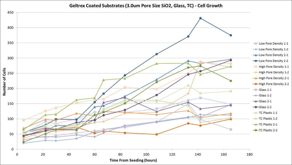
The Geltrex coated substrates, however, do have fairly strong viability:
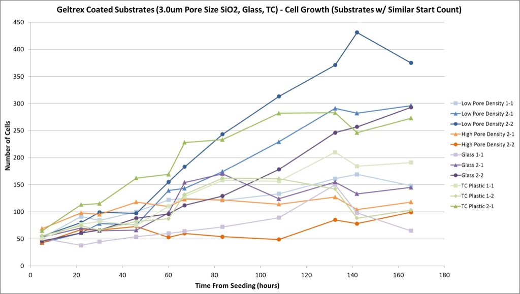
I also took a sampling of substrates which started at a similar cell count (40~70 cells), to see how they compare to each other. It does appear that glass and TC are fairly similar, and that the lower density 3.0µm pores allowed for strong growth also (even surpassing the standard surfaces to some extent):
The second trial yielded similar data in terms of coating conditions. Both the Collagen coated and non-coated surfaces seemed to have poor ability to sustain the HUVECs:

*The Low Pore Density membrane was broken from the beginning, but some cells did adhere initially so I decided to count to see if they would be viable (they weren’t).

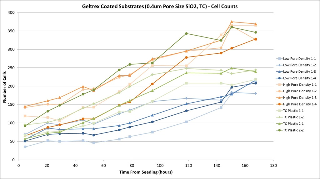
And again, the Geltrex equivalent conditions have much great viability and proliferation of the cells:
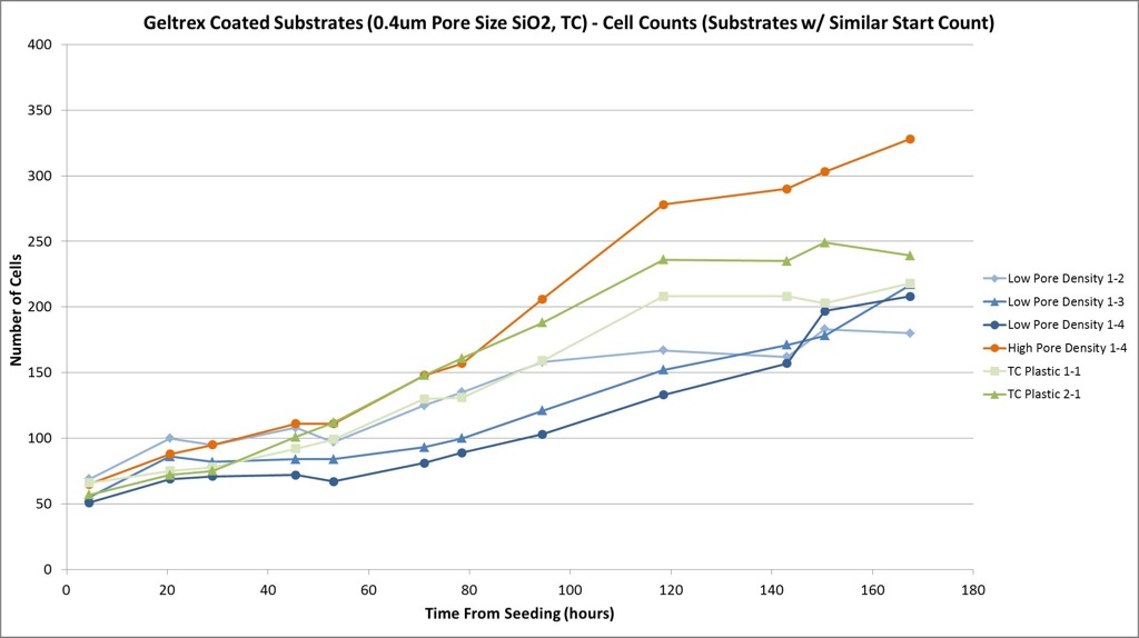
When comparing the similarly started cell counts (~40-70), it appears that the higher pore density of 0.4µm pores allowed for stronger growth, though lower density still offered growth similar to TC:
From this data, it seems a Geltrex coating provides for the best growth conditions overall (and prevents cell death and lack of proliferation). The 0.4µm pores seem to favor more growth when at a higher density, while the 3.0µm pores do so at lower density. The opposite densities do still provide for some level of proper (if slightly less) growth as compared to TC.