Modification of parylene C/N membranes for altered cell-substrate interactions
The motivation of this work by Daniela Lincoln (master’s student) was to investigate the feasibility of using different variants of parylene (C and N) for barrier studies and to look deeper into the implications of plasma surface modification for cell-substrate interactions.
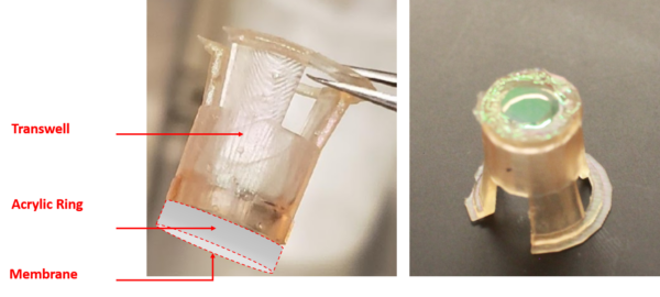
For this purpose, we used and tested a variety of device designs, which led to a 3D printed scaffold similar to transwell system (Figure 1). Originally the purpose of the acrylic ring was to provide a more smooth surface for membrane attachment, but we soon realized that it can additionally serve for easy and more convenient staining and culture. The acrylic ring has pressure-sensitive adhesive on both sides, one side attaching to the membrane and the other part attached to the scaffold. By using a tweezer and with enough carefulness, the ring with the membrane can be detached at any step of the cell culture and staining process, be it for easier culture on the bottom side or for easier staining.
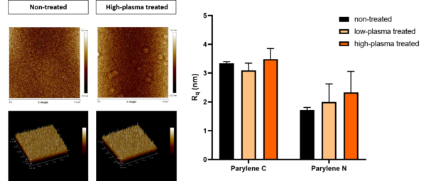
Using this system, we are aiming to characterize the physical implication of plasma treatment. Figure 2 shows the results of the limited number of samples we have run so far. It seems that there is a trend of increase in roughness as a result of increasing treatments, but we have to wait for more runs to draw a meaningful conclusion.
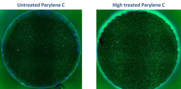
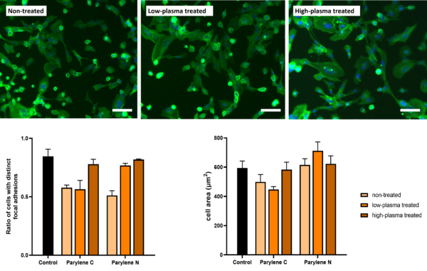
At the same time, we are trying to use hCMEC/D3 BBB cells and HUVECs to investigate how such treatment or even the switch from Parylene C to N might have implications for altered cell behavior. We aim to characterize this through analyzing cell attachment ratio to seeded cells, cell spreading, focal adhesion formation, and fibronectin/ECM deposition. Figure 3 shows a stitched image of representative samples of treated and non-treated samples, stained for nuclei and cytoskeleton. Figure 4 demonstrates the preliminary data for cell spreading and the ratio of cells which distinct focal adhesions.
Building upon this initial data, we aim to do a more thorough characterization of focal adhesion and fibronectin deposition and extend these experiments to HUVECs (already started and seeded). We also will look into cell behavior in sparse and confluent monolayer conditions.