Images of 1st BBB co-culture (+ 2 extras)
This post presents cell images from the first successful co-culture experiment. I seeded P8 bEnd3 cells alone, P13 NG108-15 cells alone and both cells on PET and SC500 pnc-Si transwells. For this experiment, I seeded 50,000 bEnd3/cm2 on the apical side and 50000 NG108-15/cm2 on the basolateral side of the transwell. I tracked TEER (future post) and stained the samples with Live/Dead after 14 days of culture.
On PET:
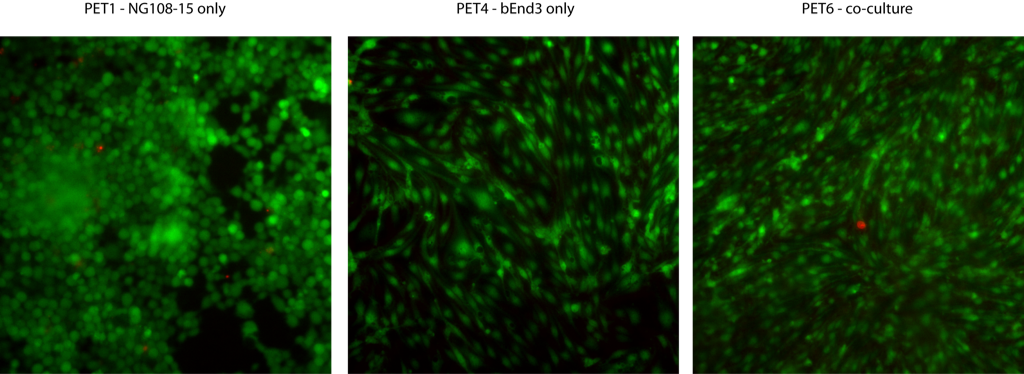
To get these images, I stained the NG108-15 cells only in the basolateral well and found near-confluence (left). Since these cells are poorly adherent, I likely sheared some cells off during staining/rinsing. The bEnd3 monolayer alone exhibits ‘tyical’ morphology (center) – here I added Live/Dead to the apical well only. Interestingly, the bEnd3 cells in the co-culture (right) expressed no vacuoles – here I only stained the bEnd3 cells to minimize fluorescent signal from the NG108-15 cells.
On pnc-Si:

The left and center panels are pnc-Si transwells with bEnd3 cells only. Since these cells were on the apical side, so you can only visualizecells through the pnc-Si window. As expected, these cells expressed many vacuoles and some complex 3D/tubular structure. The right image is the NG108-15 cells alone on the basolateral side of the transwell. Even though these cells are hanging upside down, they’ve filled up the well of the pnc-Si transwell.
pncSi co-culture:

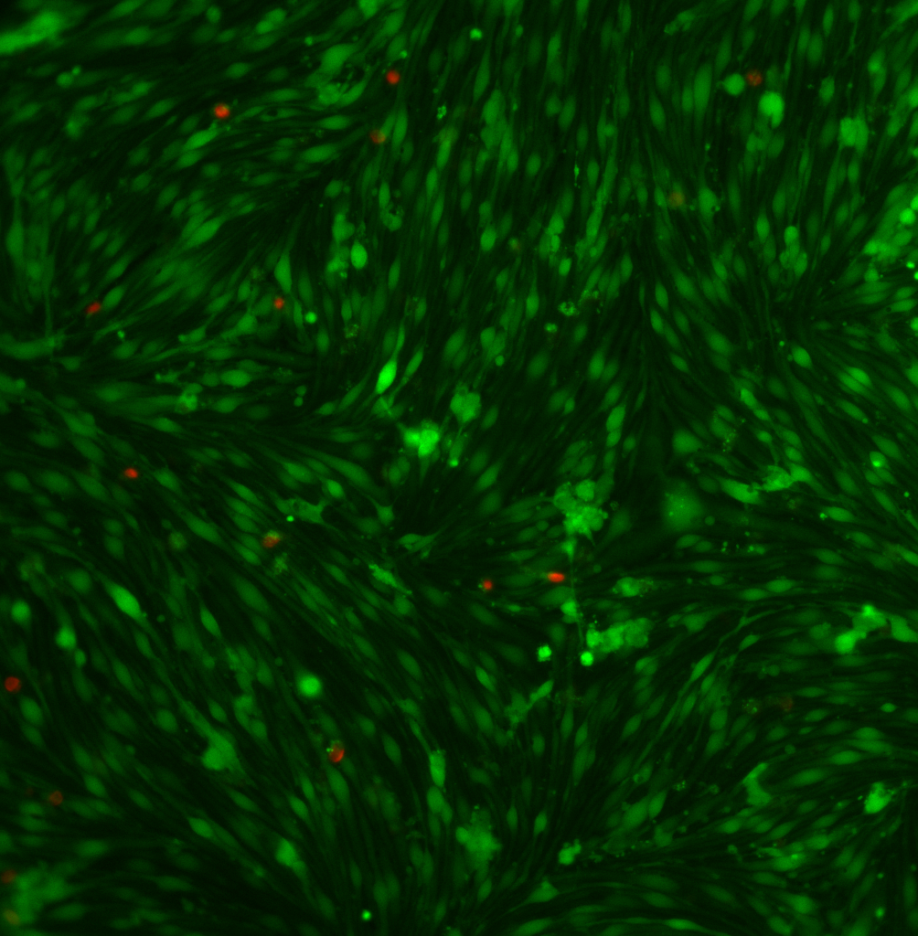
To get the left image, I stained the bEnd3 side only. The image was blurry (and the NG108-15 cells didn’t stain anyway), so I stripped the NG108-15 cells off the bottom and re-imaged this sample (center). Th cells look worse than those of the monoculture – few vacuoles, atypical morphology and low density. The right image is phase contrast and you can clearly see large, round clumps of NG108-15 cells with the bEnd3 monolayer underneath.
This experiment shows a difference in endothelial cell morphology between mono- and co-cultures. Unfortunately, the bEnd3 cells look weird in the co-culture. Since the endothelial cells are the barrier cells of this model, I am most interested in how they behave over the membrane. Therefore, I plan to repeat the co-cultures with bEnd3 cells on the basolateral side so I can stain and visualize the entire monolayer.
As a control, I cultured cells in 2 wells of the 24-well plate to test if vacuoles form on flat substrates. The image below demonstrates that vacuoles do not form on flat TCPS:
I obtained another interesting image by adding Live/Dead solution to the apical well of an SC500 transwell and allowing it to diffuse through the membrane to stain endothelial cells on the basolateral side of pnc-Si. This demonstrates the possibility of investigating intercellular signaling (i.e., Ca+2 waves, gap junction communication) by introducing dyes/effector molecules/drugs to a specific geometry of cells through a highly porous membrane and probing the speed/distance of the resulting signal.
