Electrodes: electrical measurements and EO tryouts
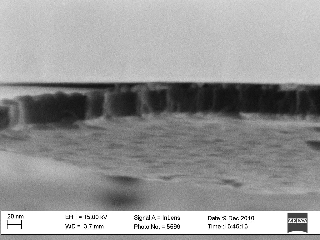
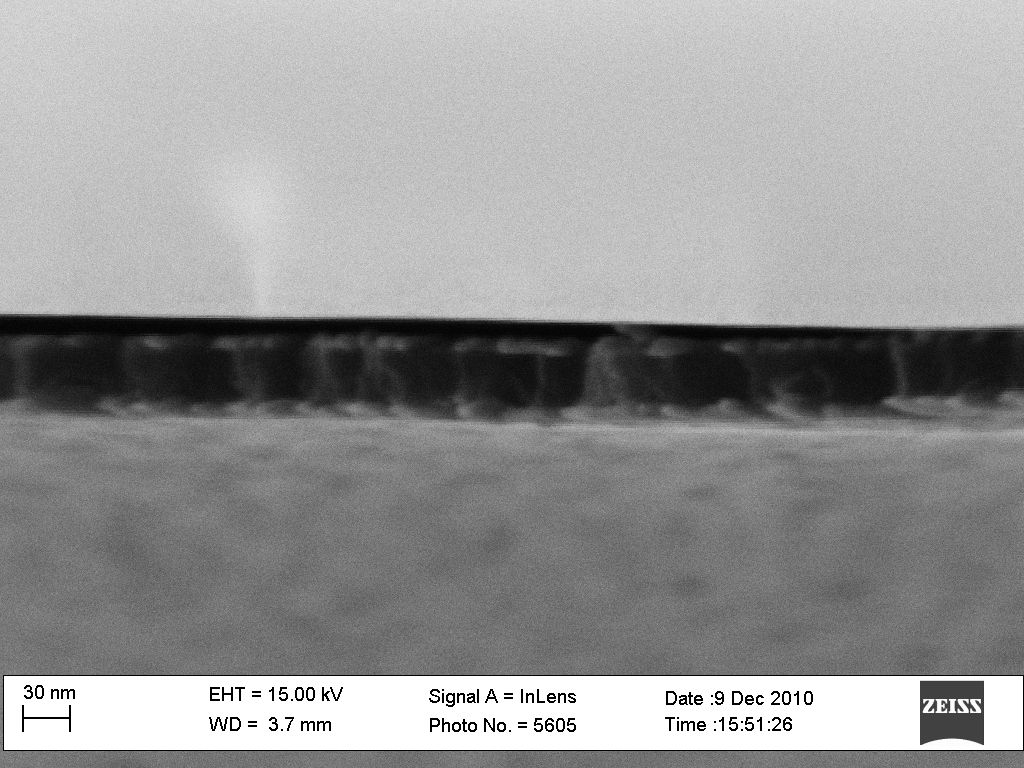
In continuation of work on ultrathin metal electrodes on pnc-Si, I tried changing metal thicknesses at different positions inside the chamber. In this experiment, on the front side of the 30 nm thick membrane the 22 nm metal layer was deposited from three different positions in the same run. The back sides of all the samples have 14 nm of metal deposited form single position. The SEM images of the depositions are below. Depending on the position in the chamber the layers are not continuous.

I checked resistances of all the layers. Three sample types have following results:
a) top surface: >MOhms
b) top surface: Ohms; bottom surface: Ohms; across two surfaces: >MOhms
c) top surface: Ohms; across two surfaces: Ohms
So the samples form the deposition B seem to have electrically separated electrodes, although the porosity dropped significantly in that run.
The cross-sectional SEM images below show cleaved sample A. Although its very hard to see the structure of the cleave is 30 nm membranes, I think they show that metals are on different sides. Samples A and B are not very different in deposition conditions so they probably look the same in cross-section.
We tried doing EO test with Jess on the samples that show electrically separated electrodes. No positive results on water flow yet. This is probably due to almost no porosity in these samples (B) after 22 nm metal deposition. I will tell more at the meeting. The attached paper describes the experimental setup that ideally I want to use EO. The image below is how we did it.