Membrane dimensions
Below is an optical image that shows the amount of area loss caused by the preferential EDP etch.
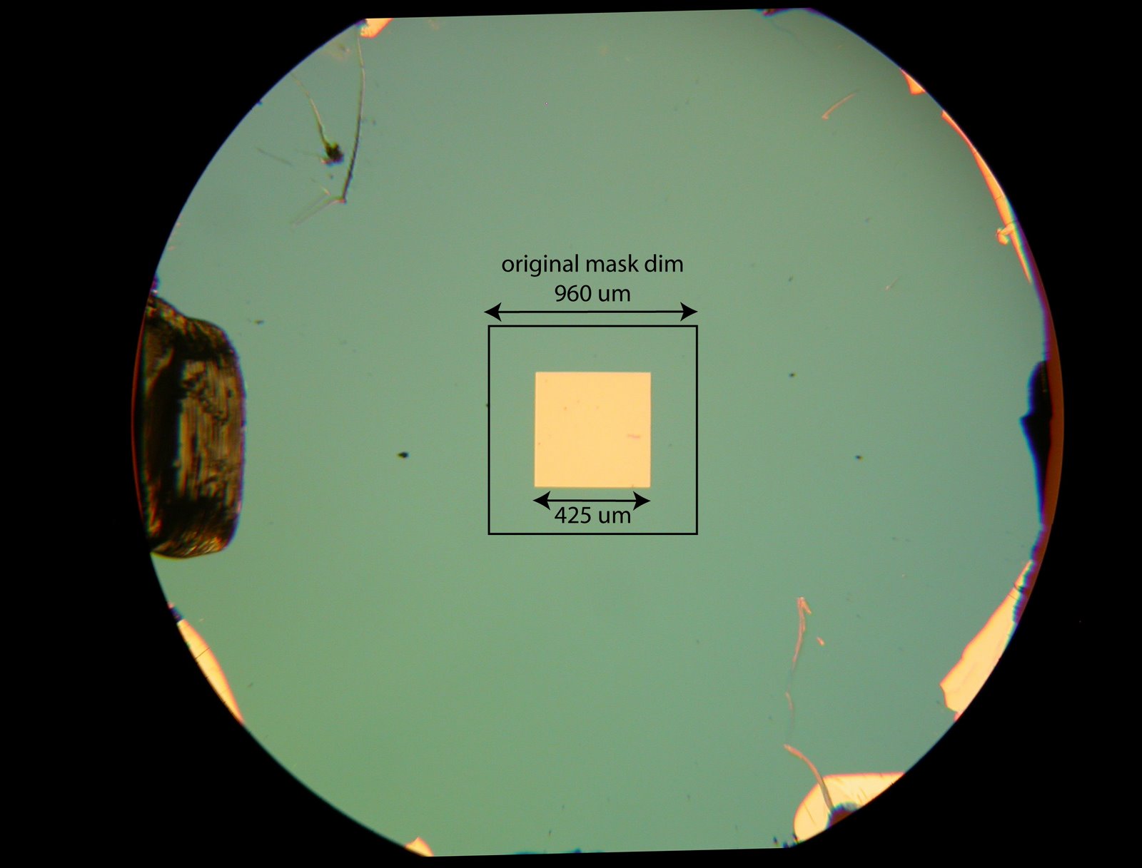
Based on these numbers, our most recent membrane slits measure 115 um x 2014 um.
Below is an optical image that shows the amount of area loss caused by the preferential EDP etch.
Based on these numbers, our most recent membrane slits measure 115 um x 2014 um.
I set out to do an in depth study of adsorption to various surfaces using the sample buffer method. Midway through staining the multiple gels I noticed there was a problem. It looked as though glass and polystyrene adsorbed about the same amount of BSA as did pnc-Si. Details of the setup follow. Preparation: Using…
For everyone conducting biological studies, this paper highlights how to think about sample size and replicates. It suggests using “SuperPlots”, which color codes data points from different experiments to better visualize the type of variability seen (i.e. random variability, experimental variability, etc…). SuperPlots: Communicating reproducibility and variability in cell biology Jim and I recommend everyone…
Katia in Dr. Shestopalov’s Lab functionalized some pnc-Si windowless chips. The process she follows is shown in Figure 1. She provided chips at various stages of the process. “Grignard”, “Diazirine”, and “PEG”. After functionalization, the chips were wetted with 20 µL of 1:1 pbs then 40 µL of 5 mg/mL f-BSA was added. The…
Good morning, We received some messages from customer who are having trouble logging into their Matlab account. If you are having trouble accessing Matlab, please see below for your update instructions. If you continue to have Matlab issues, please contact Matlab support at http://www.mathworks.com/support/contact_us/index.html. License number 680214. Your activation Key is:28093-41703-14274-60467-52214. Matlab update instructions: Open…
Jamie asked me to post these images of our membranes taken by Dr. Xiao Zhang from Hitachi. They used a 300 kV tool to acquire these micrographs. The spacing between the atoms (striations) tells us the crystallographic orientation; most of the nanocrystals we see are in a (111) orientation. It’s interesting that the perimeter of…
Here’s the paper we’re discussing this week: Wu(Moulin)JMS14 The authors motivate their work by claiming that ultrafiltration membranes can be used to keep “engineered nanomaterials” (ENM) out of our waters. They use confocal laser scanning microscopy to locate the position of fluorescent CdTe quantum dots (QD) on/inside an Ultracel membrane. (Made by Millipore using regenerated…