TEM Tomography of NPN and npMgF2
Many, many thanks to Kilean Lucas and Brian McIntyre who enabled our metrology.
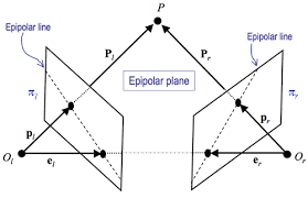
We have desired to get 3d information about the structure of our nanopores. One method to do so is known as electron tomography, a technique which can reconstruct volumes and surfaces from sets of tilted images. By identifying dense regions of contrast in TEM images, and knowing the viewing angle between image, we can triangulate fiducial marks between frames and recreate a set of slices that represent the volume of the shifted image.
Methods
I used a custom plugin for ImageJ with manuals found at this link
http://www.cmib.fr/en/download/softwares/TomoJ.html
TomoJ Tutorial – Download the Tutorial Image files from the Above Link
These reconstructions were produced from a simple weighted back projection. There are more rigorous reconstructive algorithms available that may provide superior resolution in the future (SIRT). I followed the tutorial process flow as much as possible. I occasionally downscaled images to enable faster computation (500×500 pixels).
NPN Reconstruction
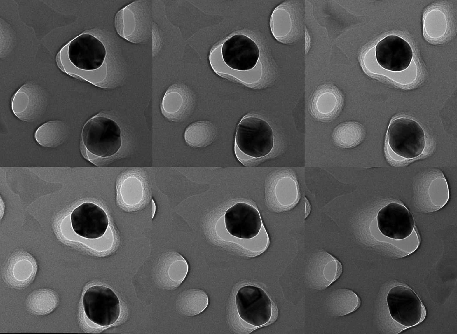
Using TopoJ, we take the tilt stack and create a tomogram, where the individual contours align with the edges of the structure. Alternatively explained, the tilt images are a guide to our perception of where edges are in 3D, and the tomogram takes that guide and reslices the stack so the edges are where they should be.
Here we see the contours of the conical pores, as well as the gold nanoparticle.
MgF2 Reconstruction
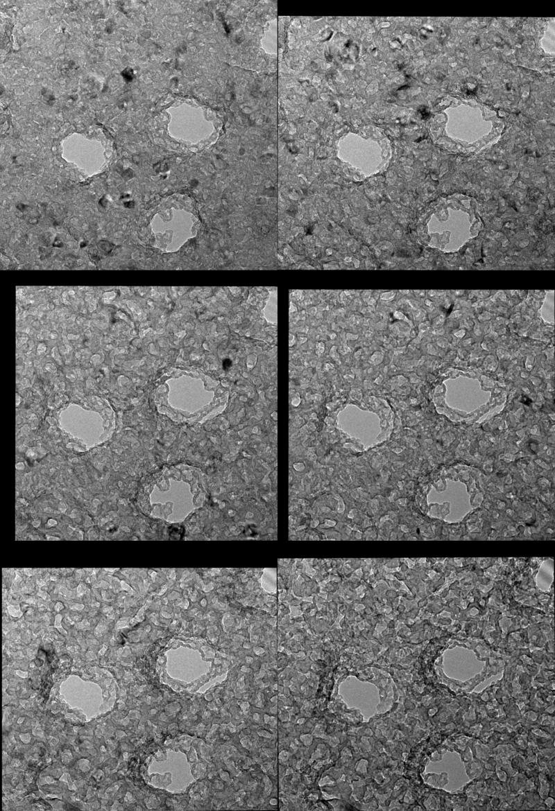
Using TopoJ, we take the tilt stack and create a tomogram, where the individual contours align with the edges of the structure. Alternatively explained, the tilt images are a guide to our perception of where edges are in 3D, and the tomogram takes that guide and reslices the stack so the edges are where they should be.
The above movie shows the MgF2 Volcano 1081E Tomogram. Starts at top surface and moves through to the bottom face. The pores are narrowest at the top of the volcano, and then broaden to where they would be open on the flat side of the nanomembrane
In the future:
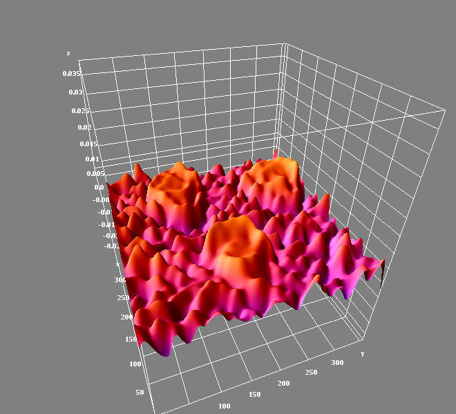
With appropriate thresholding and identification of structures (Segmentation), we can create a much more realistic volume. If each volume is thresholded so that the solid parts volume are white, we can more easily visualize the interesting features.