HUVEC adhesion – high cell seeding densities
This was my first experiment in which the Sepcons were inverted for cell adhesion. After allowing the cells to attach for 4.5 hours, I inverted the Sepcons in media for staining with CMFDA. Therefore, the Sepcon is set up so that the pnc-Si side is down and the cells are “upside down”. This makes the microscopy very easy.
Based on some of my observations with TEER, I have been using much higher inital seeding densities than Anant. My original goal for this exeriment was to determine if very high cell seeding densitied caused TEER values to increase more rapidly than lower seeding densities. After discussing with Anant, I decided just to look at how close to confluence these densities set up the cells.
I compared Sepcons to Corning PET transwells. Cell densities were low (43,750 cells/insert) or high (87,500 cells/insert). These values are equivalent to ~130,000 cells/cm2 (low) and ~265,000 cells/cm2 (high).
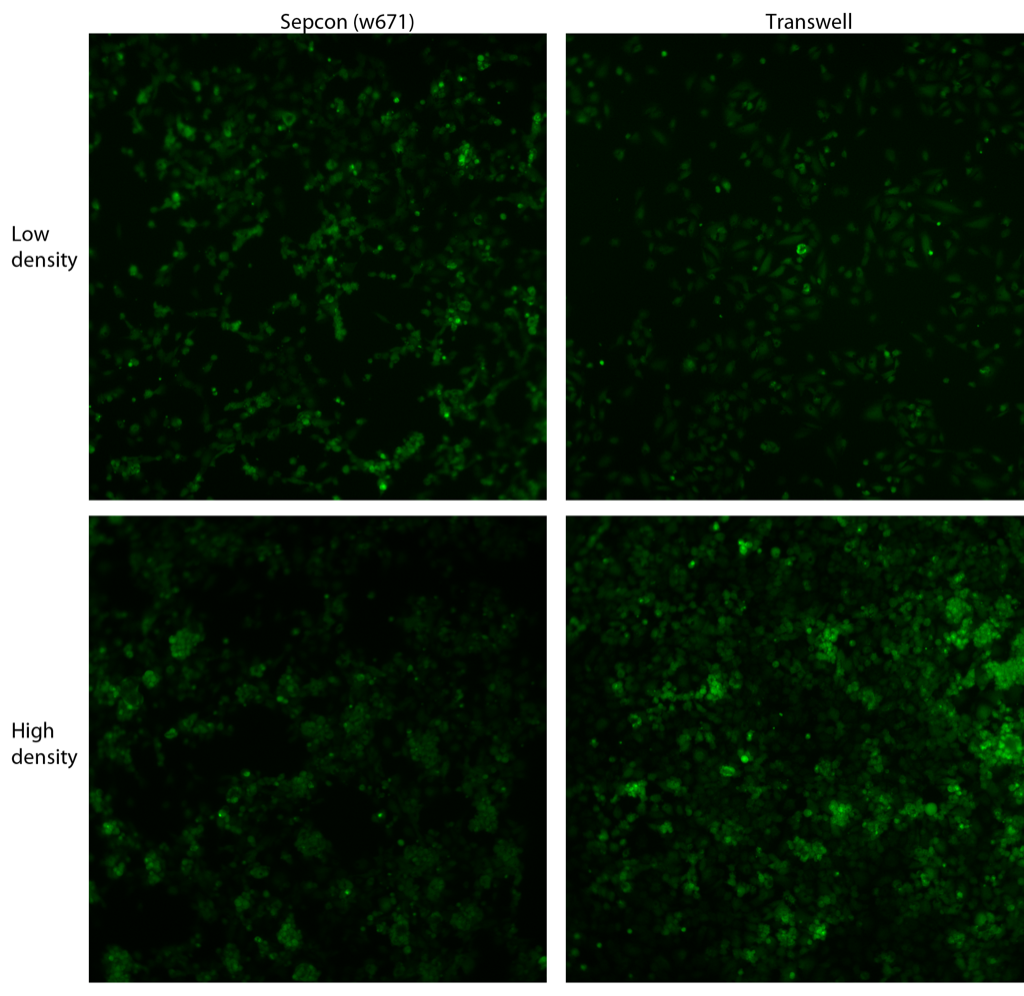
Both of these densities caused a nearly confluent layer of cells to adhere to the substrates. So many cells attached that I couldn’t run the cell counter without having to manually adjust more than half the counted objects. The high density allowed multiple layers of cells to attach, which is undesirable for monolayer studies. It looks like the low density is about the highest possible that can be used while still maintain a monolayer after adhesion. Therefore, I think we should go higher than Anant’s 11-15k cells/cm2 but stay well below ~100k cells/cm2 in future studies with transwells.
Interestingly, HUVEC morphology on pnc-Si looks better than in recent posts but it looks really good in transwells compared to pnc-Si. Qualitatively, cell adhesion percentages look about the same between PET transwells and pnc-Si transwells.
The cells on the pnc-Si/low density picture that are in the background look fine except that they are out-of-focus. The darn clumps of cells are in focus however and give the bad ‘morphology.’