Andor Dragonfly Whole Membrane Imaging
This is just a quick comparison of the new types of imaging that we now have available to us. As Bill mentioned previously, we have an Andor Dragonfly confocal system now available to us thanks to Patrick Oakes. We have been testing the system extensively and I have been messing around with it for imaging nanoparticles at the whole membrane scale.
The first thing that I did with this system was to use two different beads (50 nm far red silica nanoparticles and 100 nm TetraSpeck 4 color nanoparticles) captured on the same membrane to try and differentiate them. With the Andor system, we have the capability to montage a series of images, which allows us to stitch together a region of the chip to image a larger area than a single field of view. Well, me being me, Bill helped me to set up a montage of the whole membrane area. This is the result:
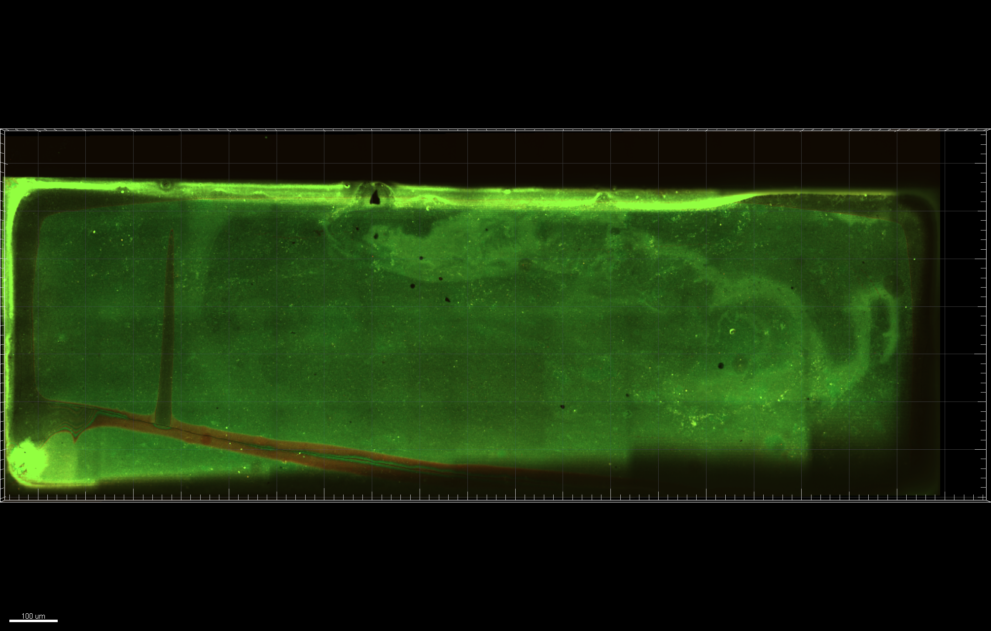
We can zoom in and start to see more individual particles:
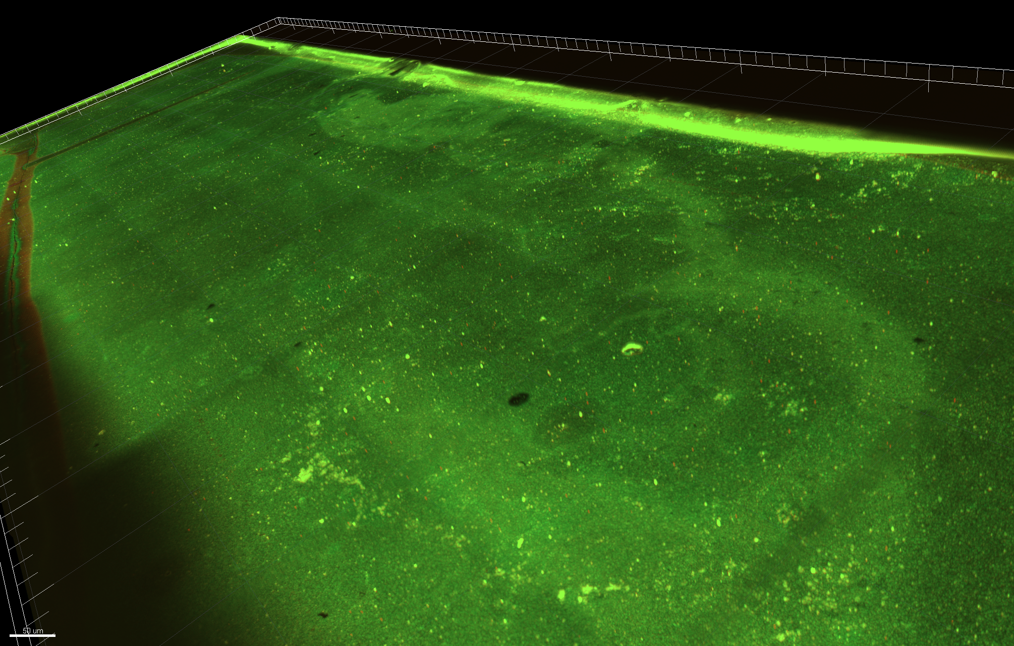
And like in the movies, because this is a high resolution image, I can say “Enhance!” and it will work:

Allowing us to differentiate these low frequency far red particles that were captured on the membrane. Furthermore, using the powerful post-processing software Imaris, we can look at a zy-plane cut:
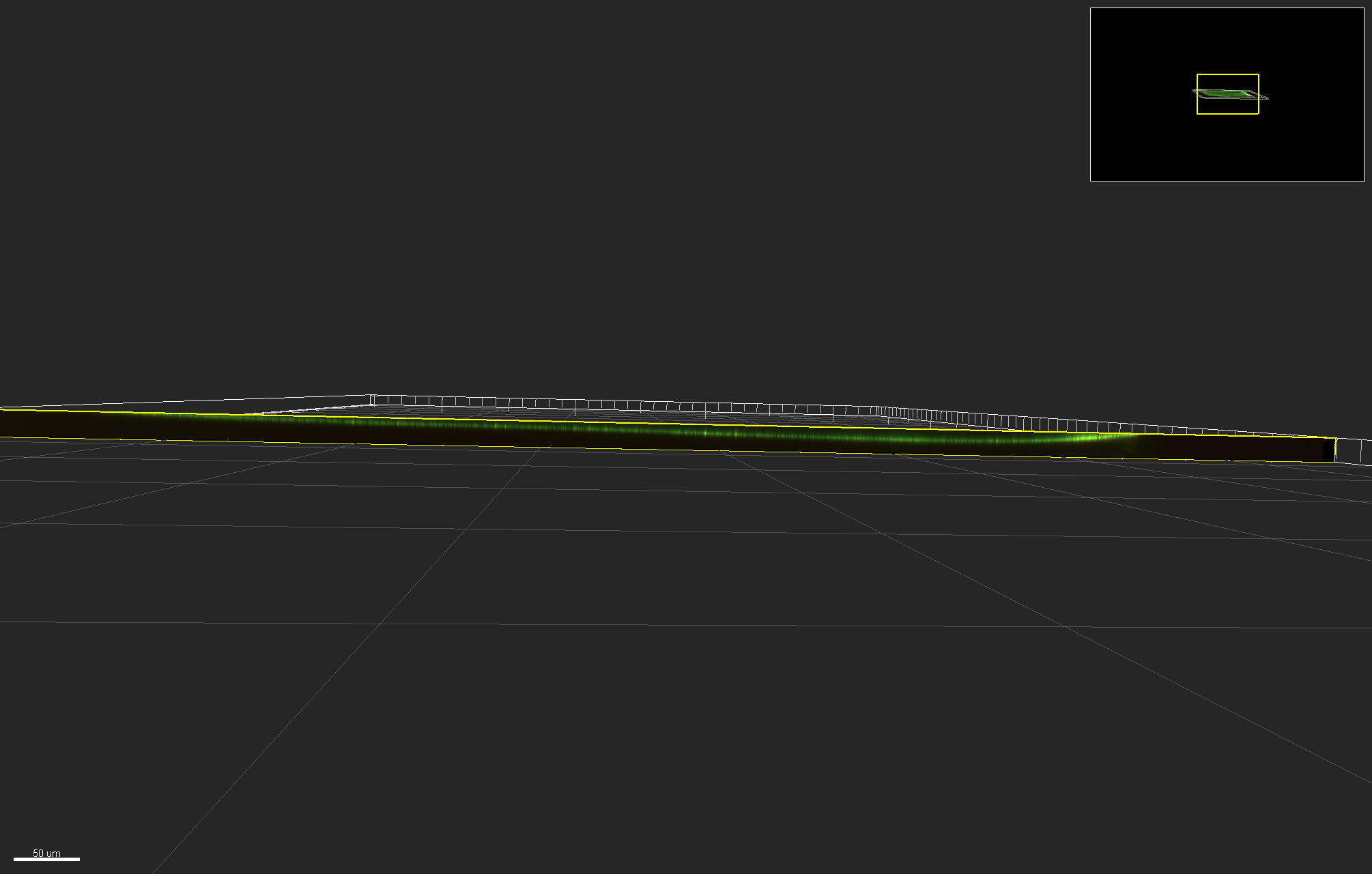
Allowing us to have some pretty sweet images. But wait, there’s more! I even made a video scanning the surface of the membrane!
Show video here. Yes, I realize that I left this in, it’s on purpose.
To demonstrate the true power of this imaging, here’s an image of a single well of a 96 well plate. What is remarkable about this image is that We can see the whole well, but furthermore we can zoom in and see a crazy amount of detail.
Show image here. Again, I did this on purpose, the image itself is nearly 200 MB to show what I want to show.