Direct Capture and Analysis of Microplastic Particulate from Environmental Sources Using Silicon Nanomembranes: Some Highlights
Microplastic contamination in drinking water is a contemporary problem that has captured the public eye, and we’re now seeing a growing demand for scientific examination of the issue. In response to this, we have recently been working on establishing a microplastics capture and analysis platform with silicon nanomembranes for environmental water samples. We started by looking at our water supply and tracing the path water takes from processing at Hemlock Lake to the tap water we drink. 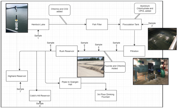
As you can see we’ve gathered 8 samples from various points along the journey water takes from Hemlock Lake. Our work process is as follows:
1. Filter water samples in a robust and scalable manner.
2. Digest organic materials on the membrane surface without adding additional contaminants.
3. Stain the membrane surface for a plastic binding fluorescent dye.
4. Analyze images for microplastics.
To be brief, we accomplished step number 1 with a gravity fed dead-end filtration setup. Glass cylinders had holes drilled into them at the bottom to allow for a measured amount of fluid to move through. Fully assembled sepcons with 8-9 µm slit pores were affixed to the bottom of these cylinders with pressure sensitive adhesive. Note that the membranes have three windows in them. Fluid samples could then be loaded into the cylinders and filter through the sepcon, leaving us with debris and residue on the membrane surface itself.
Step number 2 is done with a sodium dodecyl sulfate (SDS) digestion protocol involving 2-mercaptoethanol and tris hydrochloride. We can load sepcons into 1.5ml eppendorf tubes and use these to control digestions by ensuring any fluid from this process drains through the membrane itself. Hot water washes are utilized as well in order to remove any residue.
Step 3 is done via staining with diluted nile red dye (1 µg/ml) suspended in 200 proof ethanol.
Step 4 is currently being evaluated. We’ve seen some promising results from fluorescent imaging and energy dispersive x-ray spectroscopy (EDS)
For now, here are some highlights from the process thus far.
Post filtration: Fully processed water coming out of Hemlock Lake.
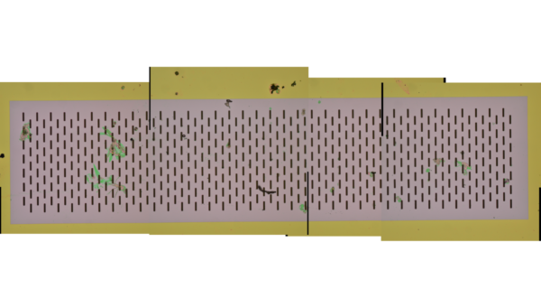
After digestion of the same window we get:
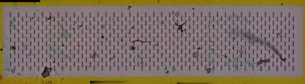
It’s a little messier, but we should note that particles move around in the digestions process and aggregates can break down into smaller components. Despite what we see on the membrane window, this water is extremely clean. After being exported, water makes its way over to the Goergen Hall where we see more troubling visuals.
Here’s what Goergen Halls water looks like from the pipes inside the building, post filtration:
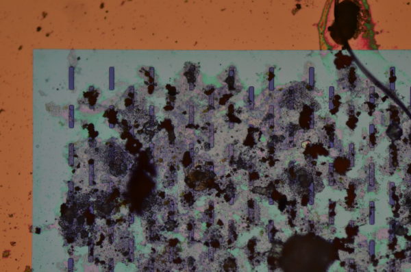
Post digestion: (Note some reduction in large debris surface area)

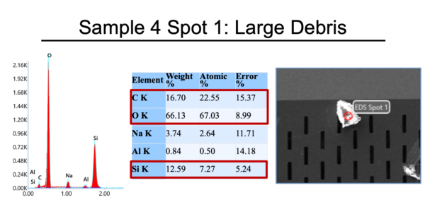
Clearly something is in the water. This issue appears to stem from water transport, but what exactly are we seeing? We can turn to EDS to see if we can learn anything about the material composition of debris in our samples.
Some highlights: Clean membrane.
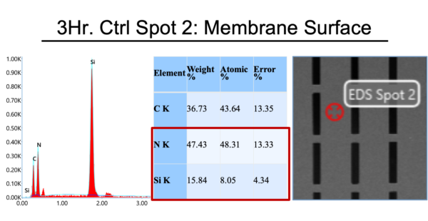
Sanity check: Membranes are made of Si and N, great!
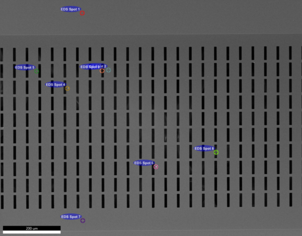
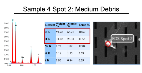
On sample four we highlighted some regions of interest. We found debris that could be plastic as well as other pieces that had different elemental compositions.
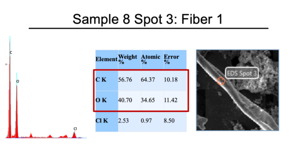
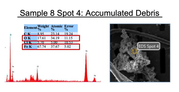
 Sample 8 had curious prospects:
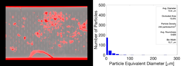
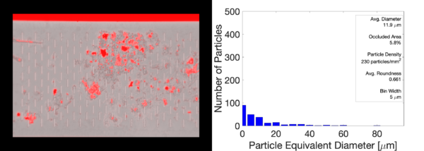
Staining can help us understand even more about what we’ve captured on the membrane surface. Here are nile red stains of membranes 4 and 8 respectively. These images are oversaturated to show stark contrast between what was stained and what wasn’t.


Lastly some particle counting work has been done with WEKA and thresholds.
That’s it for now. For fun, here’s a full montage image of what 50 mL of Goergen hall water filtered on a membrane surface looks like!

We need the following series to kick this off: 1) clean membranes; 2) membrane after filtering perfectly clean water; 3) membrane after filtering digestion cocktail. They should all look the same. Do we have that?