HUVEC adhesion update
Couple of HUVEC adhesion trials were carried out and the results were found satisfactory as seen in the charts below.

Couple of HUVEC adhesion trials were carried out and the results were found satisfactory as seen in the charts below.
This paper parallels my efforts in creating a microfluidic device that can perform co-culturing of different cell types, allow real time phase and fluorescence imaging, and perform non-invasive TEER measurement using transparent electrodes. This is almost exactly what we aimed for; the only difference is in the type of the membrane. They are using a…
This post is from Xuan ‘Sabrina’ Pan Purpose The nanomembrane used in my experiments is expected to promote efficiency of hemodialysis (HD) for end-stage renal disease patients. The membranes, however, broke during HD for rats, and breaking membranes can lead to unwanted consequences. My task is to figure out the transmembrane pressure during dialysis under…
A couple weeks ago, I posted some pictures that showed a non-linear “dose-response” curve for NaHCO3 and discoloration in the incubator (post). I repeated this experiment at 37C in the benchtop oven. These pictures show that there was a NaHCO3 concentration dependence on the discoloration rate in the oven. In the 2nd set of…
For the past month, I have been working along with Tejas towards incorporating an ECM-like hydrogel into the back (edged side) of a pnc-Si chip. The purpose of this effort is to add another layer of complexity to Tejas’ in vitro microvasculature system, in order to study neutrophil migration across the endothelium. We first pondered…
Over the past month, I’ve posted some results of a colorimetric assay for silicic acid. Previous posts are here and here. These results showed that the assay detected silicic acid after pnc-Si and PSi degraded. However, bare silicon wafers also yielded silicic acid, according to this assay. Since pnc-Si samples have the Si well-side surface,…
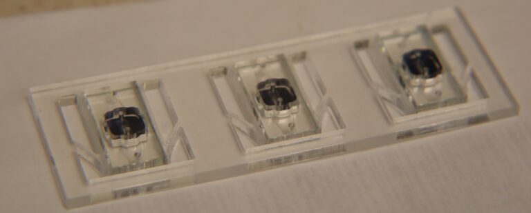
Introduction The Slide Body (Figure 1) has been used for a long time to image μSiM devices. This slide body has the same footprint as a traditional microscope slide but is designed to hold up to 2 μSim devices for imaging. The device has an open center with thin ledges where the edges of the…