HD Membrane Stability tests: 4 hour Water and PBS
This post is from Xuan ‘Sabrina’ Pan
Purpose
The nanomembrane used in my experiments is expected to promote efficiency of hemodialysis (HD) for end-stage renal disease patients. The membranes, however, broke during HD for rats, and breaking membranes can lead to unwanted consequences. My task is to figure out the transmembrane pressure during dialysis under different situations.
Experimental Setups
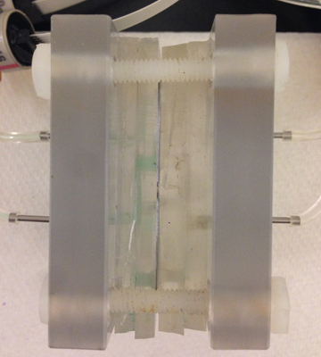
The model I used in my experiment is shown in the figure below:
The chips are clamped in between two plastic plates for fixation. There are two PDMS gels on each side of the chips. The PDMS gel that has direct contact with the plates has drilled holes that allow fluid to flow through the inlet and outlet channels, and the PDMS gel that contacts the chip has grooves that distribute fluid into two chips with equal pathway length. The top part of this model circulates ‘blood’, which will be different fluids in our experiment in which water is used as a control. This ‘blood’ circulates to the chip and returs to a beaker, while the bottom part delivers dialysate. The fluid channels on the chip directly contact the top PDMS and the membrane side contacts the bottom PDMS with the gasket in between.
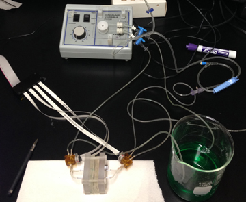
The inlets and outlets on the model were connected to four pressure transducers. The circulating of ‘blood’ was achieved using an electrical pump powered at low speed.
Method
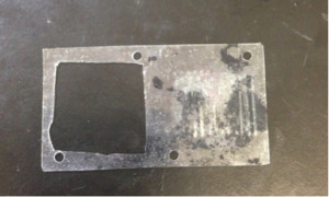
- In the first part of the experiment, a chip without a nanomembrane was used to determine the pressure difference between the side of the entering ‘blood’ and that of entering dialysate with only a gasket in between.
This gasket was used to cover the chip without membrane on it, and a dummy chip was used to balance the thickness when they were clamped between two plates.
The experiment was conducted under four conditions: the dialysate bag hung at high position and low position, and the pump working at fast and slow speeds. For each combination of dialysate bag position and pump speed, the experiment was conducted at a triplet (n=3).
- In the second part of the experiment, a chip with intact membrane was used, and this time the gasket covered the dummy chip. Water was used in both the ‘blood’ circulation side and the dialysate side. Burst pressure required to break the membrane was found by pinching off the return line of the ‘blood’. The ‘blood’ fluid in the beaker was colored using green food coloring, and breakage of the membrane can be visualized when green fluid is seen in the bottom plate side. This was repeated to find the burst pressure after running water for 4 hours.
- In this part PBS was used as ‘blood’ and dialysate instead of water to investigate the effects of viscosity on burst pressure. Other setups remained the same as in the previous part.
Results
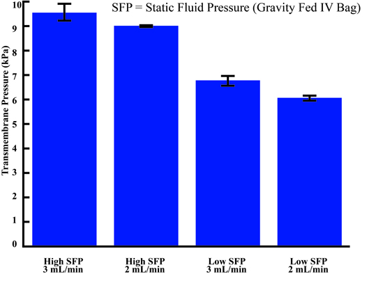 1. This figure shows the pressure difference between the inlet of the ‘blood’ circulating side and that of the dialysate. From statistical analysis, the pressure difference between high and low position of the dialysate bag is statistically significant. The pressure difference between fast and slow pump speed, however, is not significant. Greater pressure difference by hanging dialysate at a higher position can be caused by higher static fluid pressure.
2. Since the difference between fast and slow pump speed is not significant, we run the second and third part of experiment at slow speed only. The burst pressures for different fluid used are displayed in the table below.
| Fluid | Burst Pressure |
| water | ~ 11kPa (1.6 psi) |
| water (after 4 hours) | ~ 4kPa ( 0.58 psi) |
| PBS (after 4 hours) | ~ 10kPa ( 1.45 psi) |
It seems that after running the fluid for 4 hours, the membrane was much weakened, and it broke at a much lower pressure difference. But discrepancy occurred by replacing water with PBS, because a more viscous fluid is expected to have a lower burst pressure than that of using water. Even though manufacturing is different for each chip, the burst pressure for PBS after running for 4 hours seemed higher than what we have expected.
Future Directions
Following work can be determining burst pressures using more viscous fluid. The method used to find burst pressure should also be improved, since the food coloring appearing on the dialysate side will not be obvious enough to see when the break on the membrane is very tiny.