Similar Posts
Nanoporous tent wetting fluorescence inconclusive
Due to blog troubles yesterday, we have chosen to upload this post as a .pdf: Nanoporoustentwettingfluorescenceinconclusive
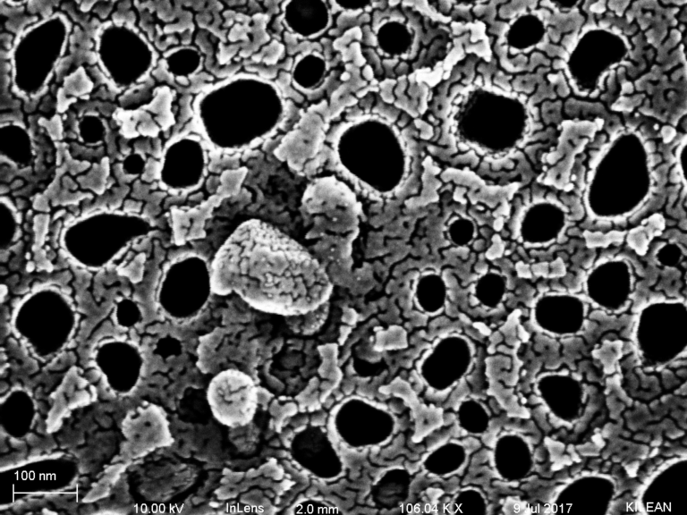
Immunogold Labeling of Exosomes
ByKileanSo, this is the data that we have all been waiting for. Without all the fancy introductions and stuff, I present you with labeled exosomes. They were isolated in tangential flow from my human plasma samples using the SOP that I have been following along with a labeling protocol that Henry and I developed. It…
HUVEC adhesion update
Couple of HUVEC adhesion trials were carried out and the results were found satisfactory as seen in the charts below.
Viability and growth of HUVECs on nanonitride membranes – Part 2
This is in continuation to the studies that I published here. I used two types of membrane surfaces: nano nitride trenches (the back side of membranes) with pure nitride on the surface, and zero-second-etch nano nitride with a completely intact pncSi cap on the top. These two surfaces basically act as materials with two extreme…
Neither Centrifugation, Sonication, nor Filtration Fix the Aggregation in the Barcikowski Lab Samples
In early May, the Barcikowski lab shipped us four samples of gold nanoparticles for us to run separations on. They took forever to get here, and had aggregated and precipitated by the time they arrived. About a week ago, the lab sent another batch of four samples (which differed slightly from the samples they sent…
BBB Device V2.6 Flow and TEER Test
Following my fabrication process from my previous post, here are the results from my flow test of BBB device v2.6: The difference in the quality of the electrodes is apparent from these images. The microscope slide electrode was difficult to spin coat, resulting in an uneven photoresist coating, and worse patterning. Etching in 12M HCl…
2 Comments
Comments are closed.
Beautiful images of aligned cells on the backside of the device! Did you take any images before flow so we can make the claim these are aligned? You mentioned that some cells didn’t survive? Can you explain where?
I might have an image of these cells before flow but it is through the clamp system.I’ll make sure to get those before and after shots when I run this for the AFM measurements. I’ll add to this post next week. The cells that are in the channel of the aline are undergoing much higher shear stresses and the cells get strip off. I noticed that the entrance side seemed to retain more cells then the exit side but maybe it is just me. The membrane is 100% confluent and healthy and I can run two Alines with same system!