Measuring Gold Separations
Even though we don’t have defect free membranes, I wanted to tweak the gold setup using membranes with pinholes. Previously I was using the small scale diffusion chamber that I use for protein separations. This setup has a retentate of 2-3 uL and a filtrate of 60 uL. To do particle sizing using the Malvern Zetasizer you need 50 uL of reasonably concentrated sample (I figure somewhere around .1 mg/mL works). The problem with the small scale setup is that you only get one chance to check the results and remove that 60 uL. Also measuring the retentate is almost impossible due to uneven dilution of the 2-3 uL on top.
I tried using the stir cell (electrophoretic cell) as a diffusion cell. Both retentate and filtrate wells hold 300 uL, enabling more samples. The following are the results from a separation using an oxidized membrane (w/ pinholes), a retentate well originally filled with stock 10 nm gold, and a filtrate well filled with DI H2O.
First lets look at the stock gold solution. I will show both intensity and number profiles. Larger particles have much higher intensity, so the intensity profile will show us if there are a couple of contaminating large particles. Number will show us the size of the particles we are looking for.
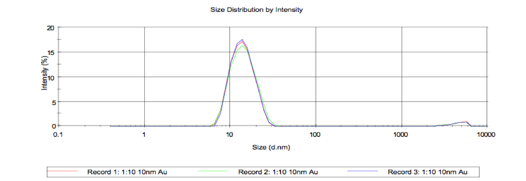
Intensity Peak ~ 14.2 nm
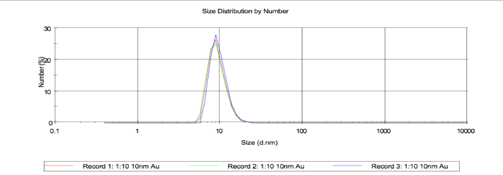
Number Peak ~ 9.2 nm
The filtrate after 1 hour:
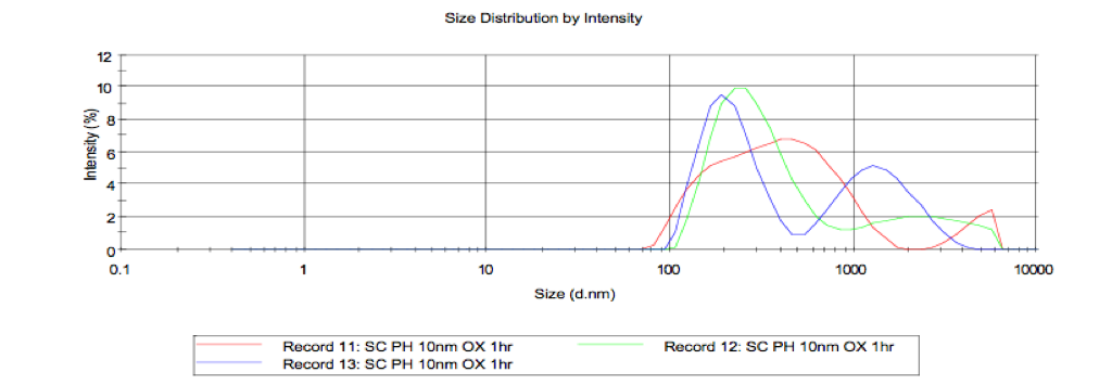
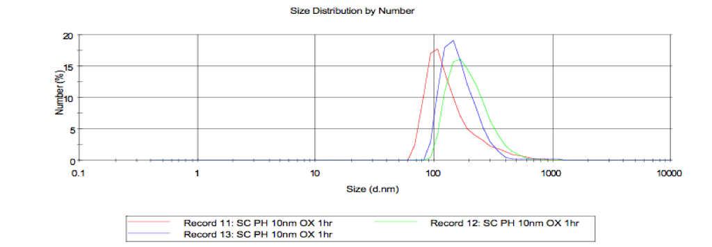
Larger contaminants are viewed in the stir cell. No 10nm gold found.
Now the filtrate after 18 hours:
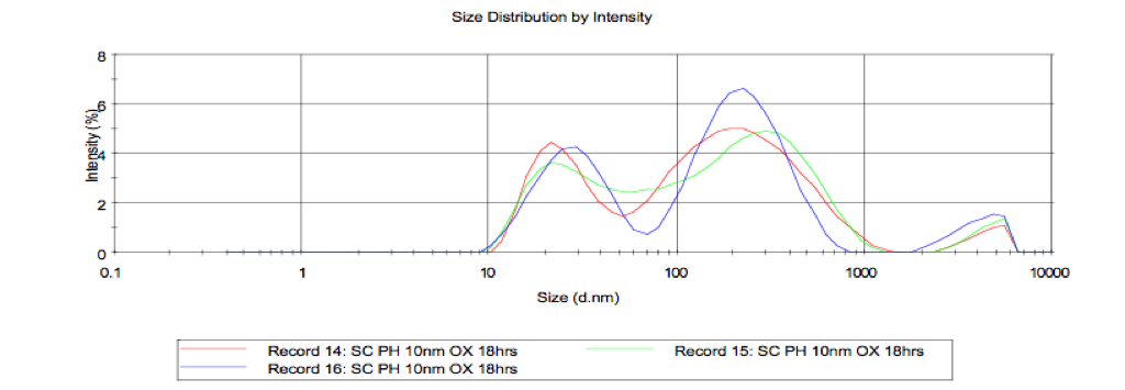
Smallest intensity peak ~ 29.4nm
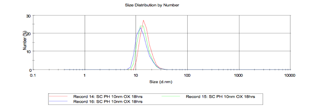
Number peak ~ 13.4 nm
Contaminants still visible, but signal from 10nm particles is strong. Number peak is higher than expected size, but this may be due to contaminating signals.
Are contaminants something being released by stir cell? Here I check the retentate after 18 hours:
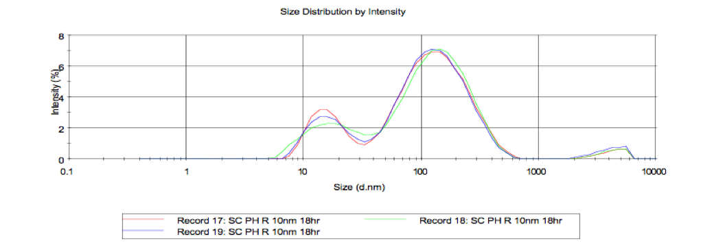
Lowest intensity peak ~ 17.2nm
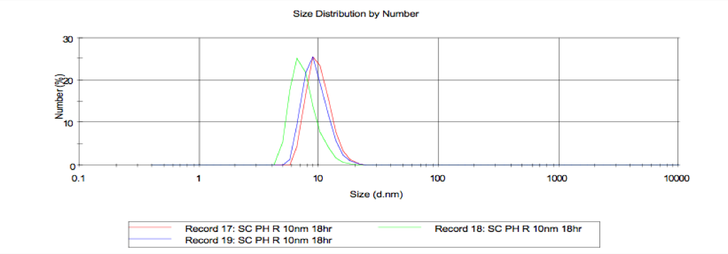
Number peak ~ 9.4nm
Larger contaminants must be due to stir cell (compare these profiles to the first ones for the stock). In this case the sizing for the 10nm particles is better than the 18 hour filtrate. This is probably because theres a very high concentration of 10nm particles as can be observed by the eye (red color).
So I think this assay seems reasonable. Whether or not this will work well with defect free membranes needs to be tested. Originally we though that separations in DI H2O were impossible due to charge interactions, but negative results could have also been caused by a bad assay. While we tried many times to modify the gold for these separations, we never tried to modify the membrane. I attempted silanization of pinhole membranes yesterday, but managed to break all the slits. I think that we will need to test oxidized & silanized defect free membranes in this new assay.