HUVECs & Microscopy with transwells
Recently, I’ve been frustrated with polycarbonate transwells since they are opaque and I couldn’t see cells on them. So, I bought new transwells from BD (Falcon, PET membrane, 0.4um pores) and decided to test these out. I also wanted to see if coating the transwells with gelatin would promote cell adhesion. These are P6 HUVECs after 1 day of culture. I stained them with 5uM CMFDA and fixed in 3.7% formaldehyde. To visualize, I placed the samples on glass slides, so PET membranes were directly on the slide but pnc-Si transwells were separated from the slide surface by the O-ring thickness. I did image processing in ImageJ – I adjusted the minimum brightness of the FITC images to the mean of the histogram.
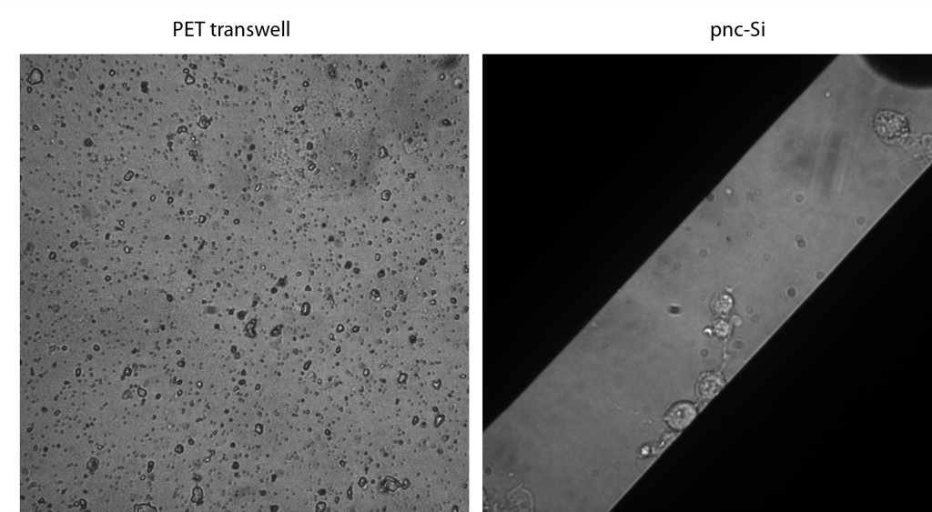
Here are 10X overlays of the phase and FITC channels. Interestingly, even though PET is transparent, there is not enough contrast to see the cells. You can pick them up after CMFDA staining, however they are totally out of focus. By contrast, you can easily see the cells in phase with the pnc-Si sample, and focussing in on them is no problem. The cells are spherical for some reason on pnc-Si – it might be due to my fixing protocol.

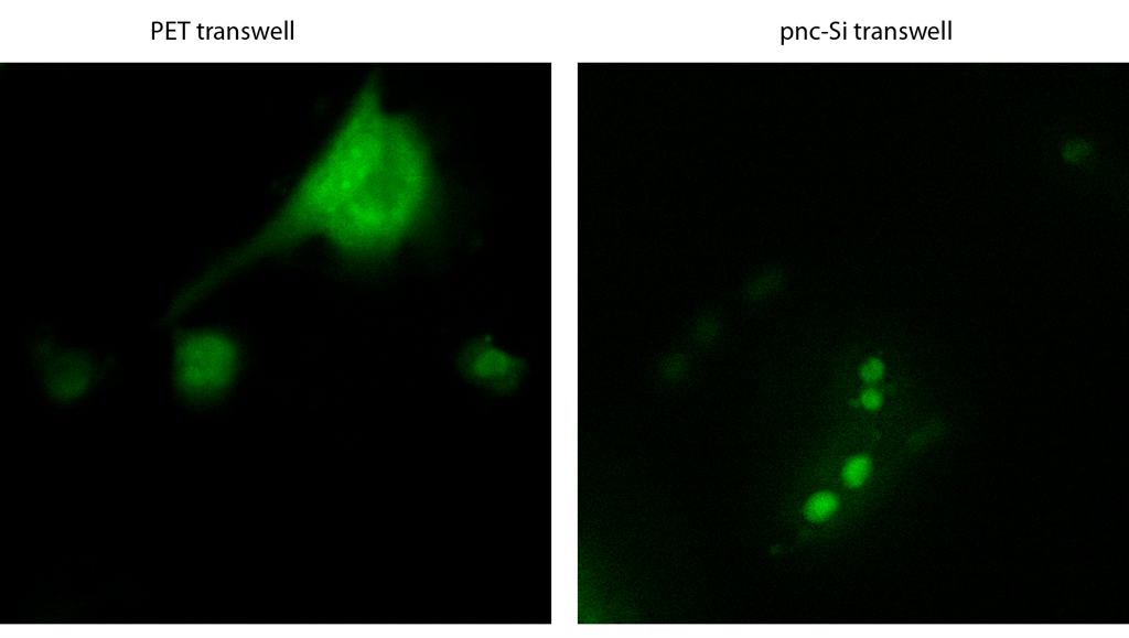
These are just the FITC channel images, which indicates the level of background fluorescence for PET and pnc-Si. There is definitely more background with PET, although it’s not horrible.

I also took some 40X images. Again, all you can see are the pores in PET transwells – no cells. The cells and intracellular detail is obvious in pnc-Si.
Here are the FITC channels from these images. The HUVEC on the PET transwell is blurry but has a somewhat normal looking morphology. I would argue that the contrast is slightly better with pnc-Si, but disappointly so. I think this might be due to the O-ring space between the objective and the pnc-Si.
It looks like the pnc-Si transwell format allows routine tracking of cell growth via phase or green fluroescent staining in 10X. These new PET transwells are worse than the Corning Costar PET transwells (see here) for phase imaging and about the same for fluorescent imaging. Any high resolution microscopy with pnc-Si transwells will require a different sample prep than I did here.