Cytotoxic Edge Effects with PET transwells?
I’ve been growing brain endothelial cells on PET and pnc-Si membrane transwells for a variety of experiments, but mainly to measure the TEER (transendothelial electrical resistance). TEER is used to track the integrity of cell monolayers grown on permeable supports. As the monolayer gets ‘tighter’, the TEER increases. PET membranes are marketed as transparent for enhanced cell imaging capabilities (compared to opaque PC) but it’s still hard to see cells on them. So, I typically grow cells on the bottom of these membranes. There seems to be a functional difference between growing cells on the bottom vs. the top side of commercial transwell membranes……..
The following is a graph which shows the %increase of TEER from day 0 (acellular) meaurements. I seeded bEnd cells @ 100,000-150,000 cells/cm2 either on the top or bottom of PET, allowed them to attach for 2 hours and then proceeded with normal cell culture. TEER was measured 1, 3 and 7 days after seeding. For cells grown on the top of PET, the TEER jumped up within a day and then stayed around that plateau level. However, when the cells were grown on the bottom of PET, the TEER increased much more slowly, only reaching top TEER values after 1 week.
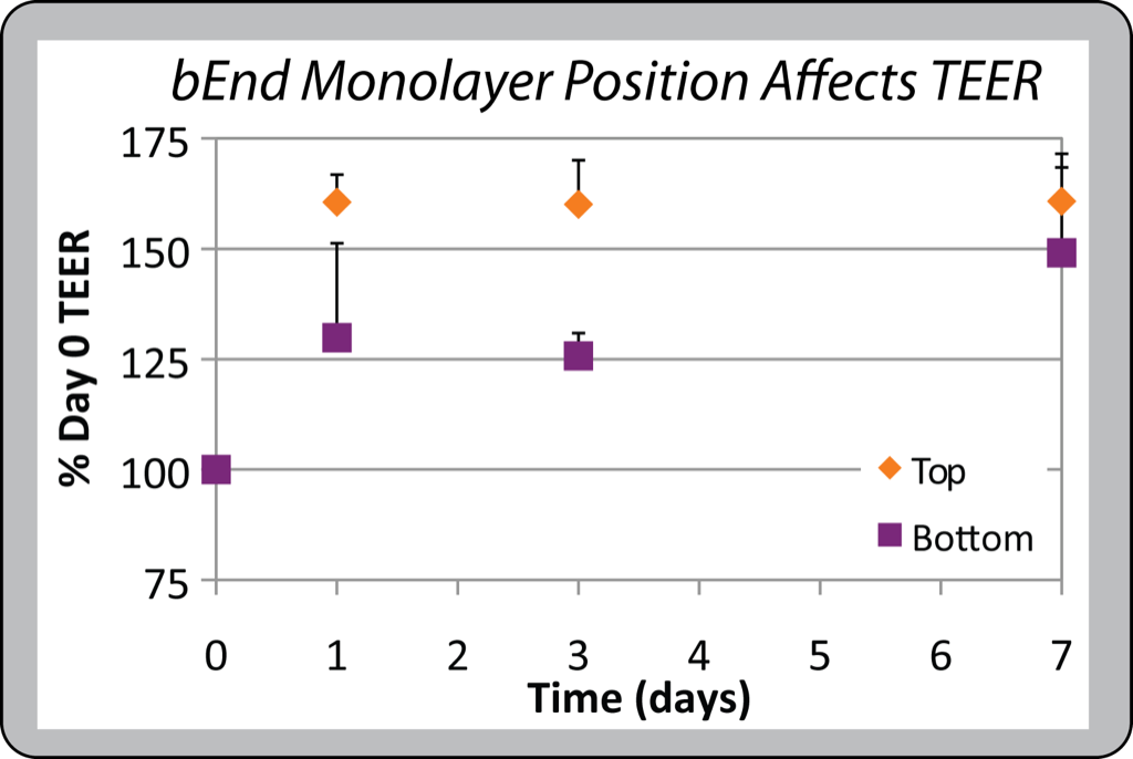
This confirmed my hunch – it’s a pain growing cells on the bottom of PET. Since you’re manipulating transwells in and out of the Endohm and 24-well plates to take TEER measurements, you have to be extremely caefuly not to knock the transwell into any surfaces or you risk damaging the cell monolayer. Our Sepcons are advantageous for this reason – the plastic and O-ring at the bottom of the Sepcon protect the pnc-Si membranes and cells from this sort of damage.
I stained cells with the Live/Dead assay to see how nice the monolayers looked after 7 days of culture.
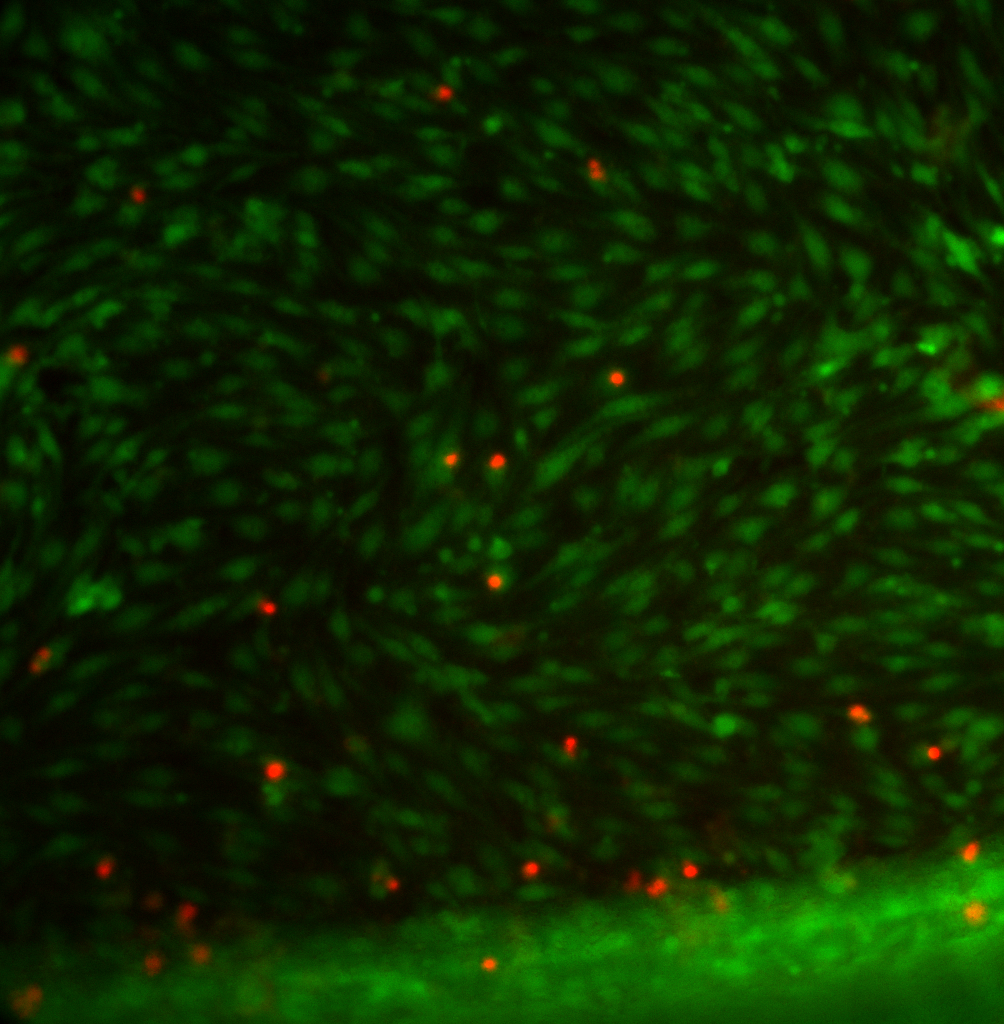
Cells growing on the top of PET membrane transwells look good – a few dead cells but a good-looking monolayer in general and cells grow right to the edge of the membrane.
20X:
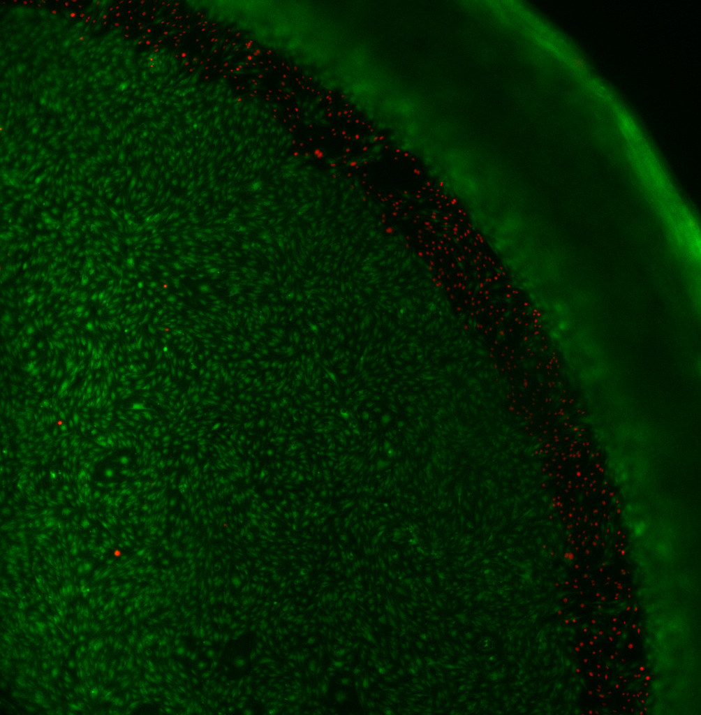
There was an annulus of dead cells on the bottom of PET membranes:
4X
20X:
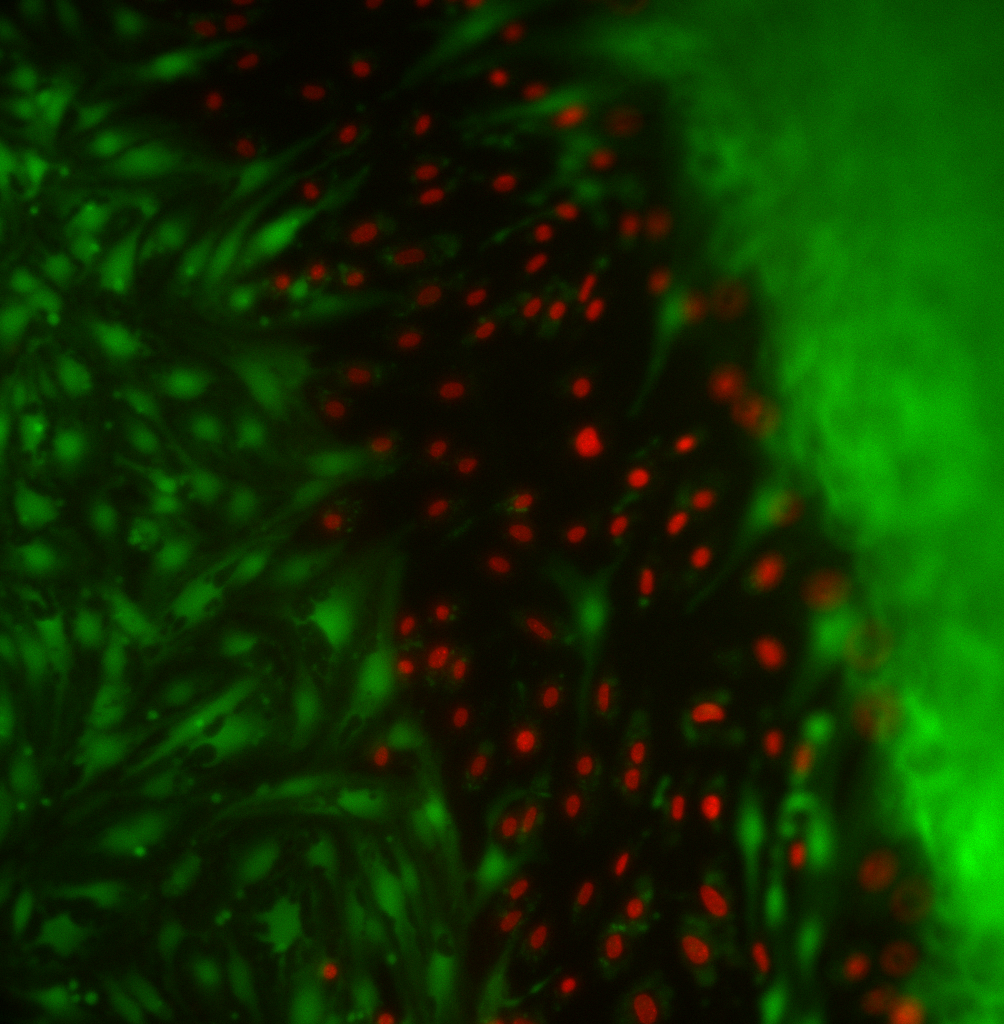
The area where these dead cells are is somewhere around where the membrane is adhered to the housing. You can see live cells on either side of this annulus and some live cells are infiltrating the dead cell layer. I showed these images to some people at Corning – they never had complaints about it and concluded that these cells were below where the membrane was bonded to the housing. This seems to be a big disadvantage to growing cells on the bottom of PET transwells – although these dead cells might not be on the freestanding membrane, I think this annulus of dead cells around the edge could cause the neighboring cells on the free-standing membrane to be less healthy (some evidence of this with the 4X image).
We don’t have to worry about edge effects since the membrane windows are in the center of chips. Here’s a 4X image of 7-day old bEnd3 cells on SC097. The membrane was intact on this 7th day – but I broke it during the staining protocol. You can see that the cells formed a similar-looking confluent monolayer as on the PET.
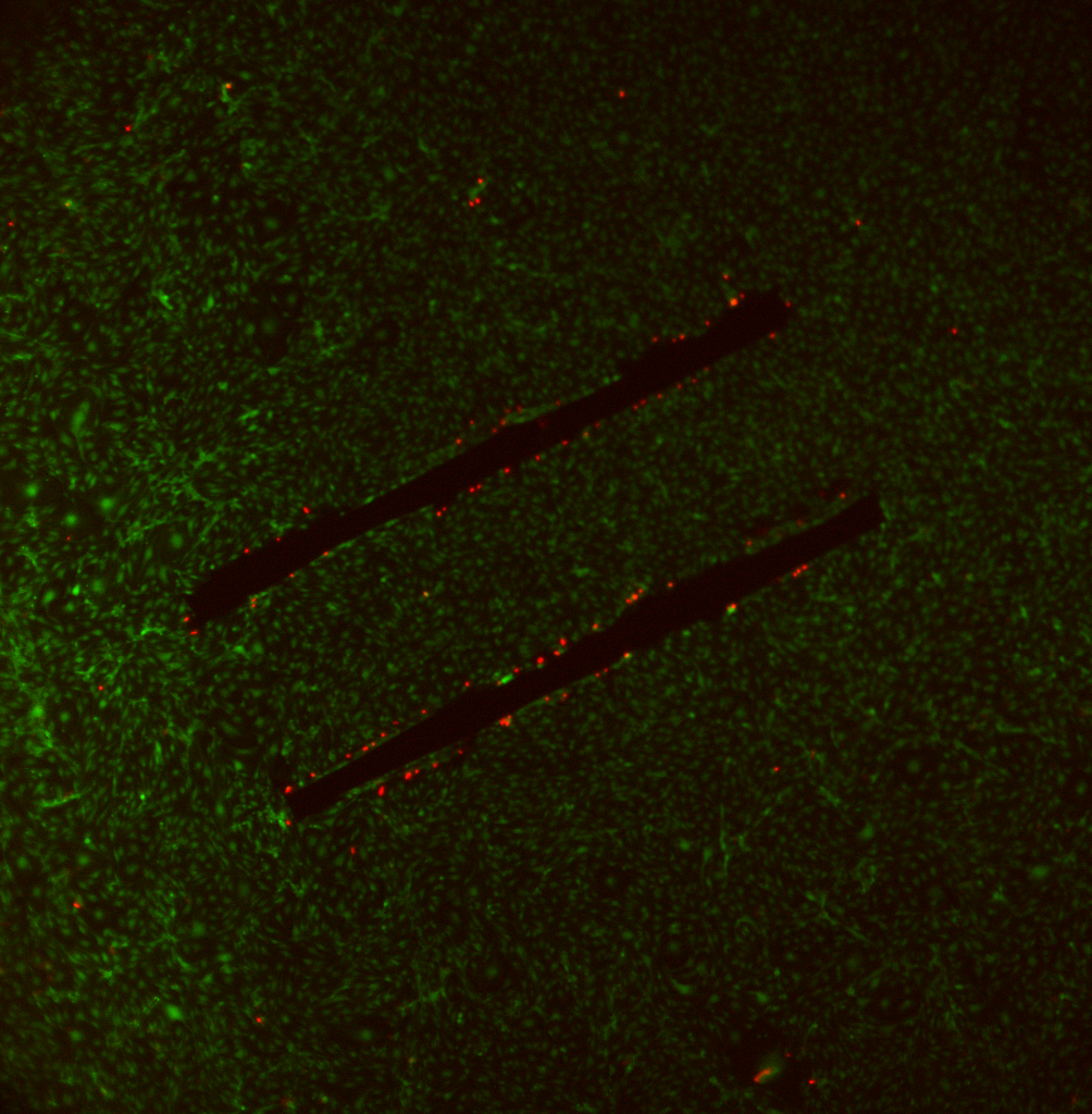
The dead cells around the membrane windows appear to be attached to flaps of the broken membrane.