TAMRA labeling of vacuoles
One of the ongoing efforts with endothelial cell culture on pnc-Si is to confirm that the holes in these cells are actually vacuoles. In my last post, I tried to do this with FITC-Dextran and TMRIA with indefinite results. I ordered some 5-6-carboxytetramethylrhodamine (TAMRA, C-300 from Invitrogen) to do this test again. This rhodamine dye should not conjugate proteins non-specifically (like the TMRIA did in the last post), and it’s the same dye used in the Nature paper I reviewed for journal club a while ago. The TAMRA dye should be pinocytosed by cells and accumulate in vacuoles over time.
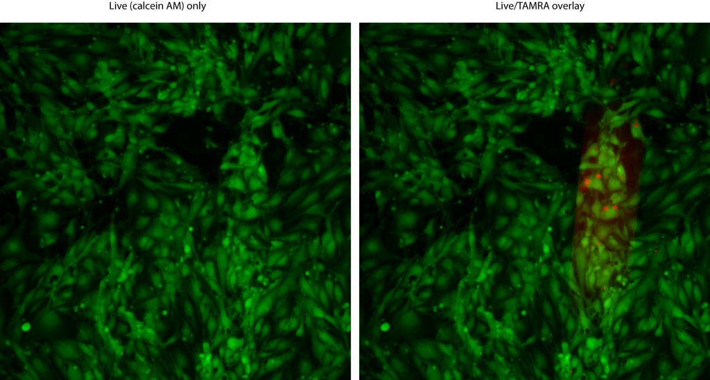
Here, I seedde 50000 cells/cm2 (P9) on SC499 transwells, let them attach for ~ 4 hours and then supplemented the media to 2 μg/mL TAMRA. I allowed the cells to grow for 24 hours in this media, then stained with Live/Dead. The following images show the green channel only on the left and the overlay on the right (TAMRA is red). The membrane window is where the ‘vacuoles’ are.
Membrane Slit 1:
Membrane Slit 2:
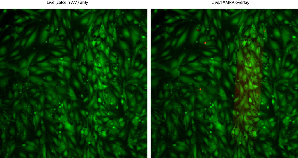
I made the mistake of using both Live and Dead stain, so some of the dead cells stained red. First note that this dye concentration is not acutely cytotoxic. On the membrane window, it appears that some of the holes were labeled with red TAMRA, especially the smaller ones. I’m not sure why more smaller, not larger, holes are labeled. It might be that the vacuoles are leaky, and thus only ‘early’ vacuoles retain the dye. It’s also possible that my labeling procedure isn’t optimized. I can try different concentrations and different exposure times to see if there’s a difference. In any case, TAMRA labeling of vacuoles seems to be working.
Great! Picture isn’t exactly what we expect yet, but still – these are indeed vacuoles! We are getting pretty close to having an interesting story to tell.