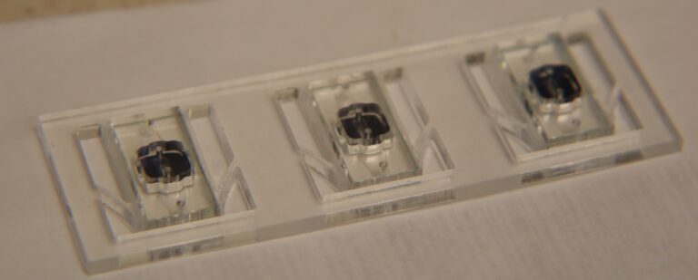
Shear-free chemotaxis: linear gradient maintained after passing through the membrane, assessed via TIRF
Hi all,
We have already showed in our earlier [ post ] that our shear-free chemotaxis system can indeed elicit chemotaxis, suggesting that we have successfully translated a gradient through the membrane to the shear-free side of the system where the cells reside. However, it is still nice to show via fluorescence how the translated gradient looks like. To this end, we imaged a translated gradient of the green fluorescent dye FITC via total internal reflection (TIRF) microscopy. TIRF was chosen because it specifically excites fluorophores within a region 140 nm off the coverslip surface. TIRF thus offer an axial resolution that is unmatched by any other fluorescent imaging modalities.
One small complication, however, is that our TIRF can only be done under the 100X magnification. We would need to image multiple times in order to entirely scan the 500um distance over which the gradient spans. We allowed 210 nm fluorescent beads to settle on the coverslip surface as landmarks/breadcrumbs to track the distance we displaced as we image to recover the spatial information.
NOTE: The source FITC gradient goes from 0 to 1 uM over a span of 500um.
Following is the combined image reconstructed from the image cross correlation of the overlapping fields of view: [combined_TIRF_image]
A line-scan of the fluorescence scanned along the mid-axis of the combined image. The superimposed red curve represents the moving averaged performed to average out the peak fluorescence singularities caused by presence of the fluorescent beads: [Image] line_scan (moving averaged).
We then performed a fast fourier transform (FFT) to interrogate the frequency spectrum of the moving averaged line-scan and removed the higher frequency components. What we kept is the first few low frequency components that describe the baseline profile of the line scan: [Image] line_scan (FFT baselined).
We then arbitrarily select a 500um segment that most likely corresponds to the gradient seen through the 500um widow width of the membrane opening: [Image] line_scan (filtered and centered). The normalized intensity difference over the 500um span is 22.3%. Assuming that diffusion never occured, the normalized difference should be 100 % over the 500 um span.
WORK IN PROGRESS: I will try to compare the normalized difference of 22.3% with the normalized difference of a gradient that averages out after diffusing 100 um away from the membrane to see if there is a close match.
CONCLUSION: We have imaged the gradient that translated through the membrane of the shear-free system via TIRF microscopy and found the gradient to be linear in shape.