Two Quick Graphs Show That BSA and Cytochrome C Both Pass Through 20nm Sepcons
Before the fresh Cytochrome C arrived, I ran two samples. The first sample was our old Cytochrome C in 0.3mM sodium phosphate buffer at a concentration of ~1mg/mL. I passed it through the 20nm cutoff 3rd generation Sepcons by spinning it in the small centrifuge at 2000rpm for 5 minutes. Approximately 100uL of sample passed through the membrane, and below is a TECAN scan of both filtrate and retentate, as well as a standard curve.

Next I ran the same experiment, but I replaced the Cytochrome C with a 1.33mg/mL BSA solution, also in sodium phosphate buffer. Only approximately 40uL of sample passed through the membrane.
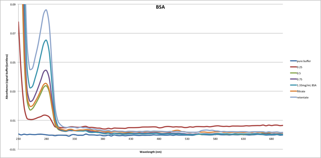
This is delightfully real data – after working at the concentration limit with the Barcikowski particles for so long, it is nice to see some unambiguous curves. Both filtrates have concentrations just over half that of their original sample, and Cytochrome C seems to be slightly more able to pass through the membrane. In this post Jess said the only time she saw separations of BSA and Cytochrome C was with a low porosity 10nm cutoff membrane. I have nearly a whole wafer of 10nm chips, and as soon as the new gasket sheets arrive (and we return from break) I will be repeating the experiment with them.