Microarray progress: slightly successful seeding and dilution/concentration variations
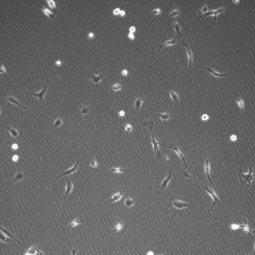
A control was done to observe cell proliferation on a TPM coated cover glass. Images were taken at 10x after 4 days.
TPM coated cover glass

Polystyrene petri dish
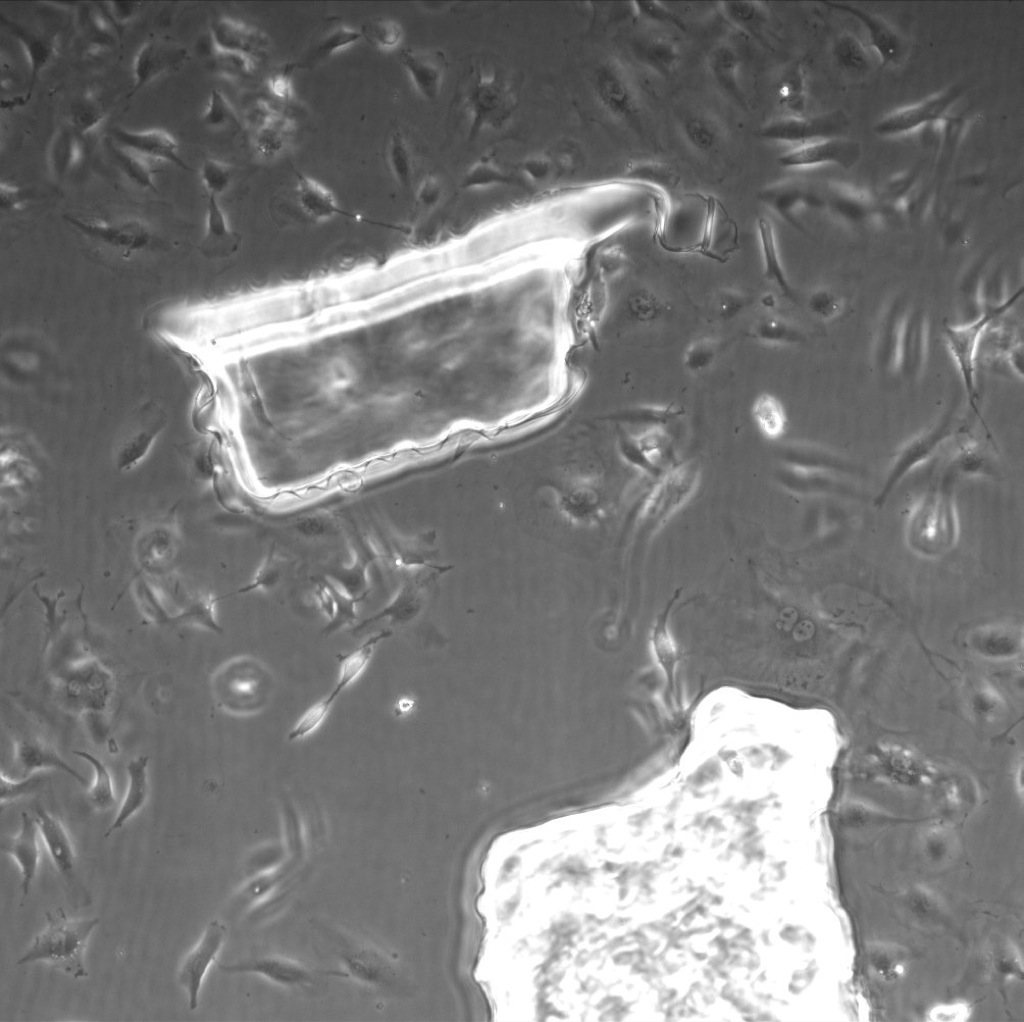
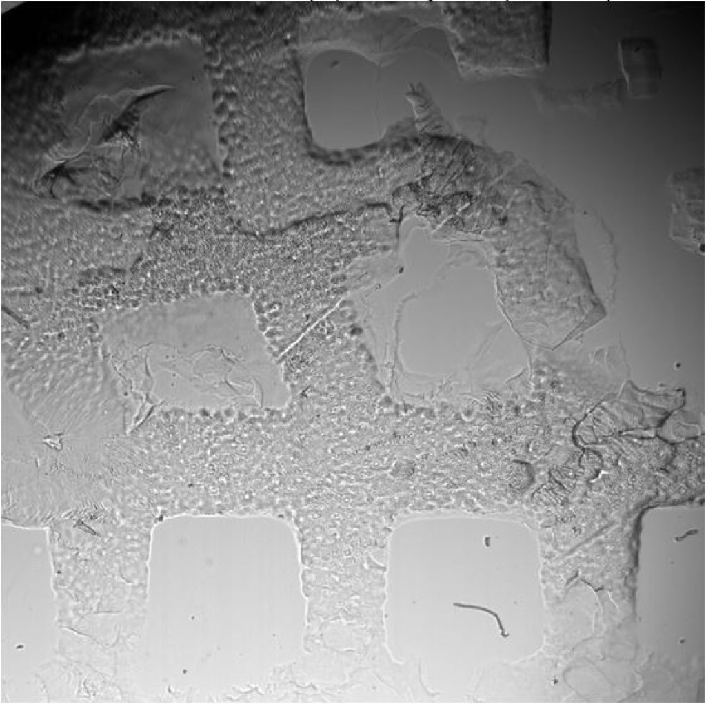
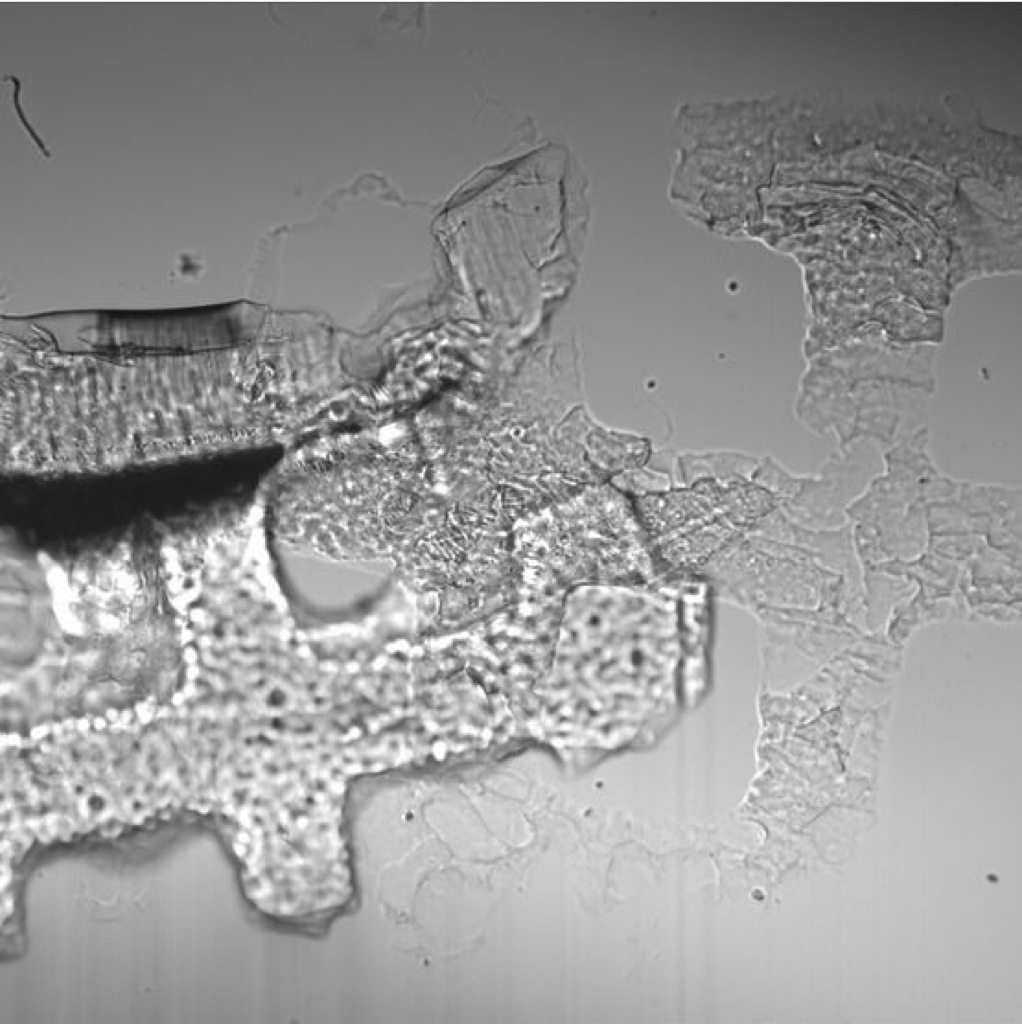
We are having some success with the seeding of bend.3 cells onto microarrays sterilized with UV exposure. These were allowed to grow for 4 days. Images were taken at 10x unless specified otherwise. Culture conditions seemed to have caused the arrays to disengage from the cover glass, but you can see that cell growth is still guided by the wells.
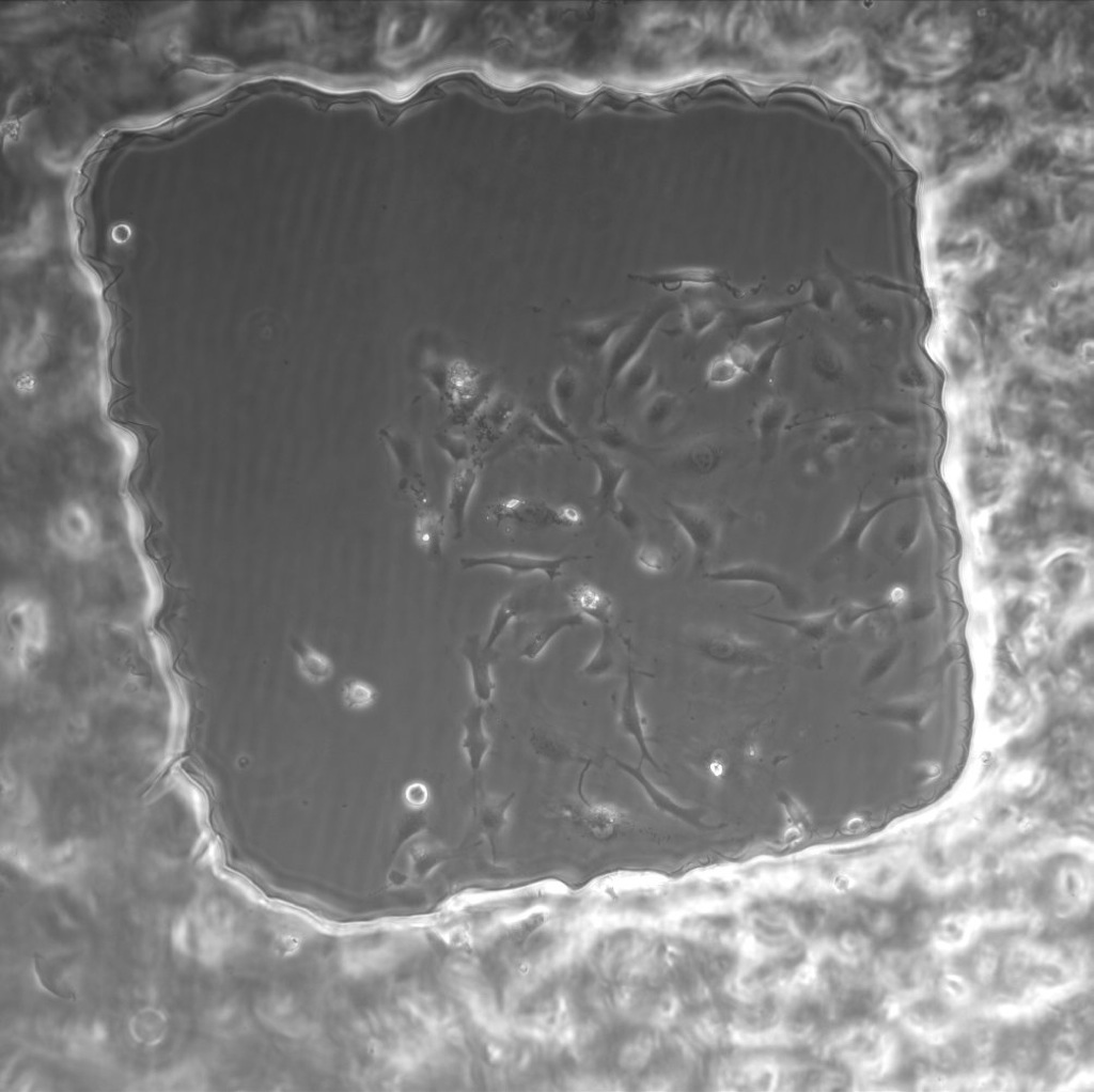
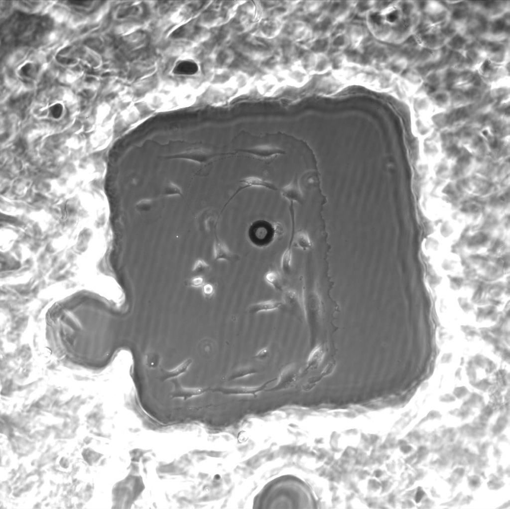
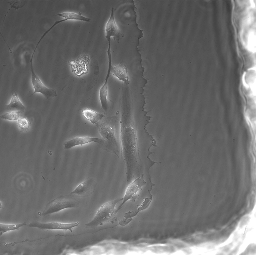
20x shown below.
This data prompted us to do an experiment where the PEG was diluted before crosslinking. We also decided to test different concentrations of photo initiator.
Varying concentrations of photo-initiator (3%, 1.5%, and 0.3%) were tested with 1:1 PEG dilutions with DI or PBS.
Exposure times:
3% – 500ms
1.5% – 2000ms
0.3% – 20000ms
Using DAPI filter cube without an objective.
0.3% did not effectively crosslink with either DI or PBS in dilution. Previous efforts to crosslink 0.3% photo-initiator in 100% PEG were successful, but I am assuming there is just not enough PEG or it to far spread apart in the dilution to effectively crosslink, even with a 20 second exposure time.
The microarrays were crosslinked on the microscope and washed once in their respective solutions. For the microarrays diluted in water, there was a second wash done in PBS to observe any effects of contraction.
All images were taken at 4x.
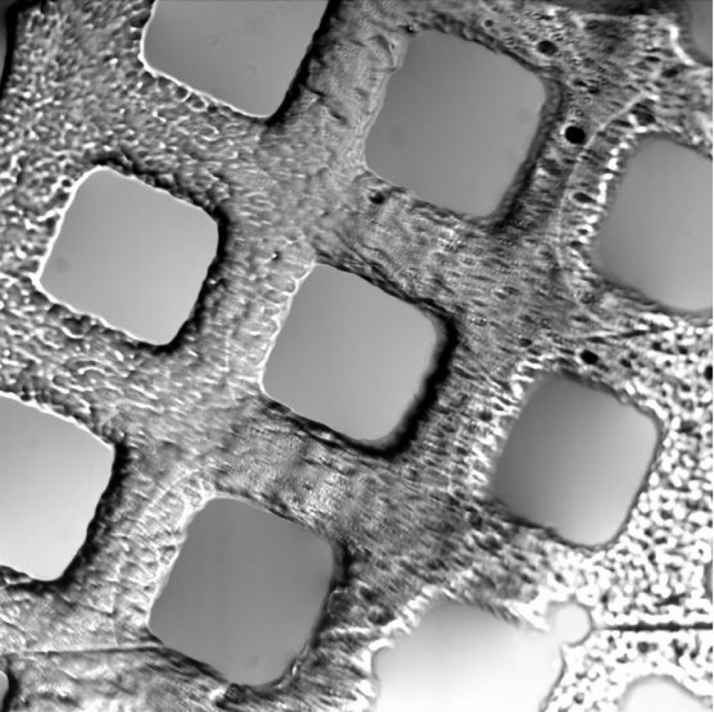
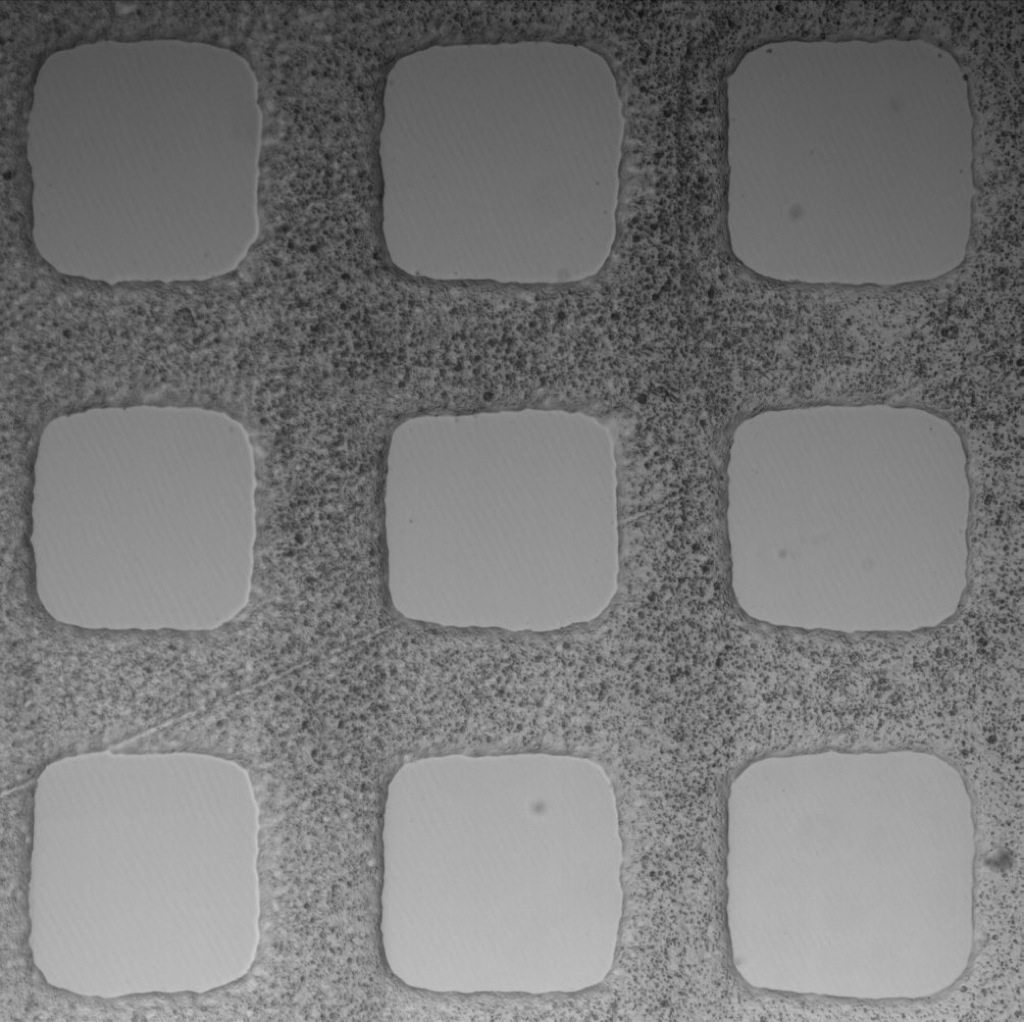
1.5% initiator in PEG+DI – first wash with DI (Maybe this was just a badly TPM’d slide?)
1.5% initiator in PEG+DI – second wash with PBS
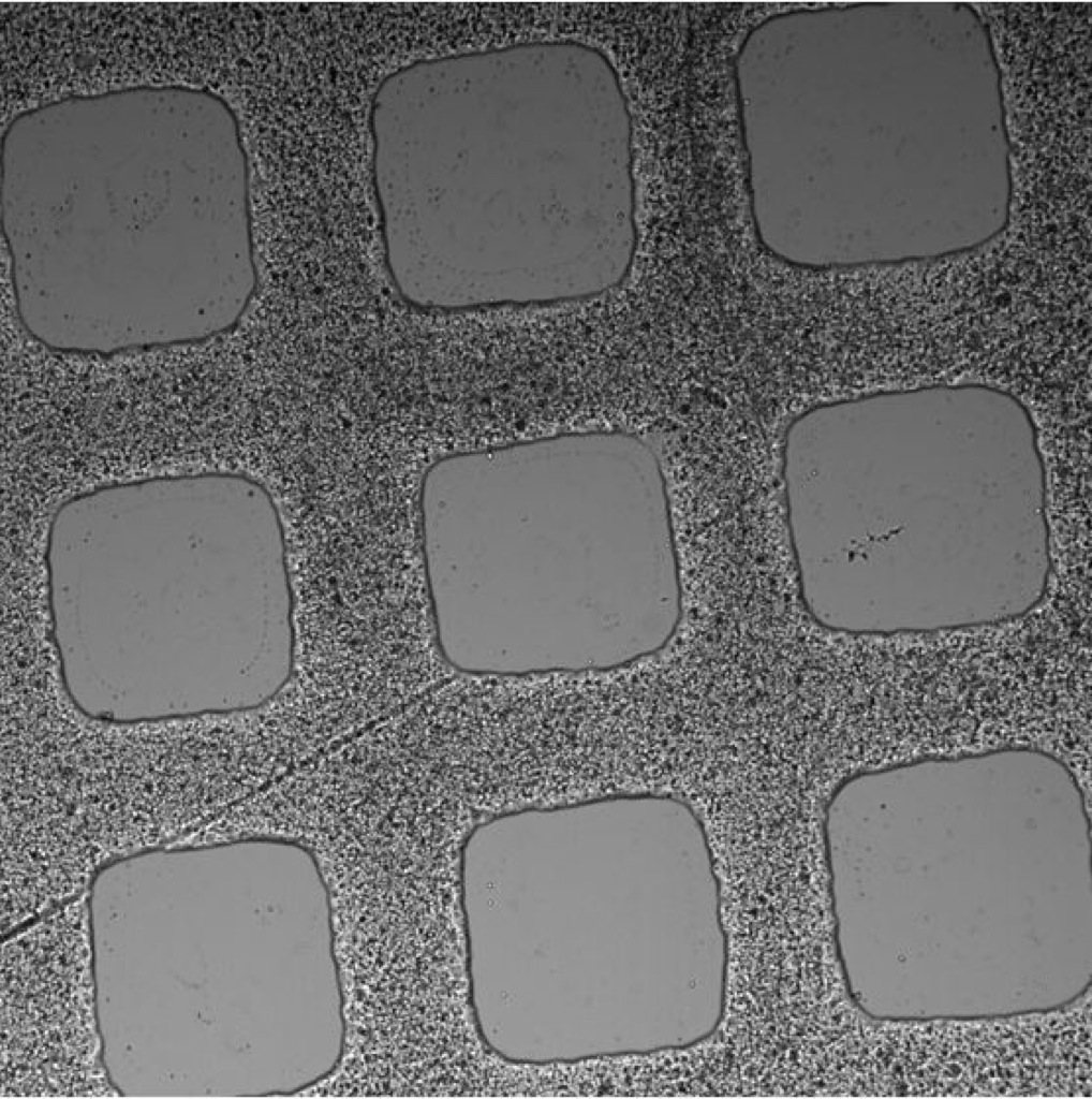
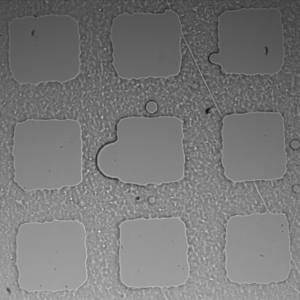
3% initiator in PEG+DI – first wash in DI
3% initiator in PEG+DI – second wash in PBS
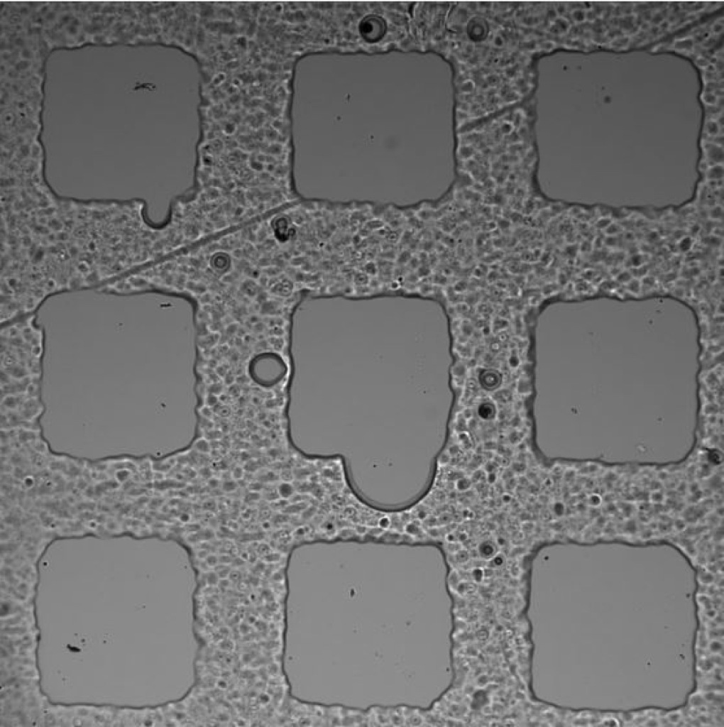
1.5% initiator in PEG+PBS
3% initiator in PEG+PBS
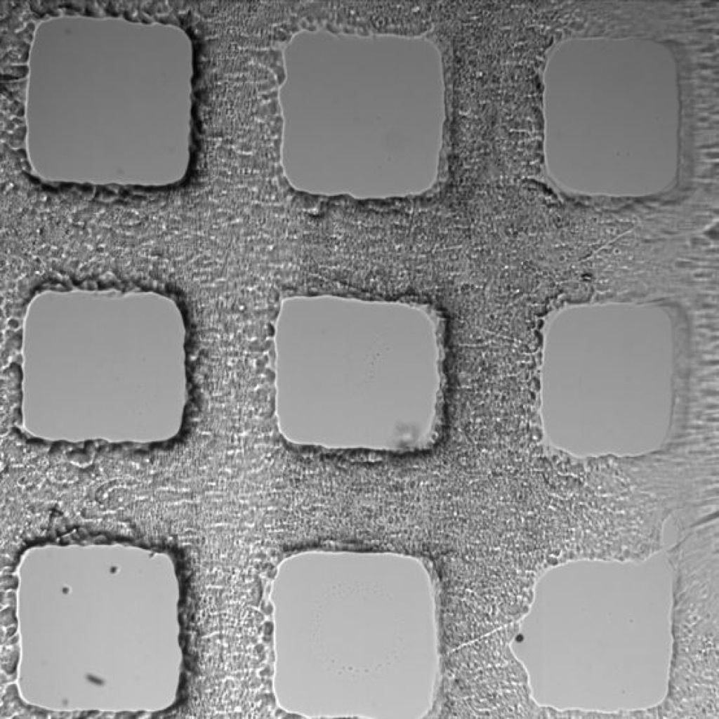
12 Days Later: Microarrays were allowed to sit in either DI or PBS for the length of time.
3% initiator in PEG+DI
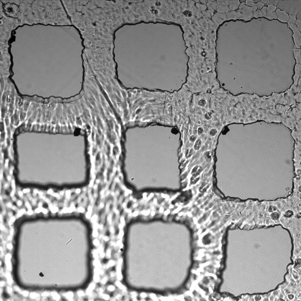
1.5% initiator in PEG+PBS
3% initiator in PEG+PBS
PBS looks like a better choice to keep the microarrays attached to the coverslips. 1.5% photoinitiator gives the most uniform features with the crosslinking conditions I being used.