BMES 2014 Talk Comments – Greg
Here are my thoughts on the talks I attended at BMES:
OP-Thurs-1-11.
BioMEMS I
Oral
Thursday, October 23, 2014
8:00 AM – 9:30 AM Room 008A
Moderator 1: James Tunnell
Moderator 2: Catherine Klapperich
8:15 AM – 8:45 AM High-throughput High-content Developmental Biology and Neurogenetics H. Lu. Abstract
- This talk used a number of image processing heuristics to separate different populations of cells based on their a) binding characteristics to antibodies or b) signal transduction from fluid stress. What was most impressive was the massive parallelism in the devices.
8:45 AM – 9:00 AM A Microdevice for Simultaneous Applications of Topographic Cues and Cyclic Tensile Strains to Live Cells Q. Wang, K. Wei, Y. Zhao. Abstract
- Much like the Lung-on-a-chip device, there is interest in cyclic loading of different cell types with different topographies at work. We can create the same types of substrates using a corona wand and PDMS. Here, they used a very fine tip on the corona wand, oxidizing a narrow strip of PDMS, which caused the membranes to wrinkle uniformly. This is much easier than using other micropatterning techniques to create the same structures, very elegant.
9:00 AM – 9:15 AM An Ultrathin Flexible Carbon Nanotube Microelectrode Array for Neural Recording and Stimulation W. Yi, Z. Feng, C. Zhou, J. Cavanaugh, C. Chen, M. Cheng. Abstract
- Another process driven talk. They take advantage of silicon processing to create a double-lift-off process for grown nanotubes, using parylene as an effective insulator/glue holding the nanostructured membranes together. They were still active. The electrodes can behave well, but there were questions about the overall stability of the process afterwards. Apparently you can etch the parylene to expose the little microelectrode heads of the nanotubes.
OP-Thurs-2-11.
BioMEMS II
Oral
Thursday, October 23, 2014
2:00 PM – 3:30 PM Room 008A
Moderator 1: Rafael Davalos
Moderator 2: Erkin Seker
2:00 PM – 2:15 PM Track Etched Magnetic Micropores to Efficiently Sort Rare Pathogens from Large Volume, Unprocessed Clinical and Environmental Samples M. Muluneh, W. Shang, D. Issadore. Abstract
- This work reminds me of Karl’s work somewhat. Metallizing a micropore can mean that we can actively control what goes through the pore. In this case, they conjugated magnetic nanoparticles to what they wanted to separate out, and then stuck a film on top of the track etched membranes. When the magnetic field is applied, the waste makes it through the large pores, but traps the magnetic nanoparticles and whatever they are stuck on. Later, they recover their treasure by turning off the magnetic field and flowing buffer through the device. We could do the same thing tomorrow with our lithographically defined devices.
2:15 PM – 2:30 PM Multiplexed Free-standing Nanowire Transistor Bioprobe for Intracellular Recording: A General Fabrication Strategy Q. Qing, L. Xu, Z. Jiang, L. Mai. Abstract
- This was really cool! They could control the growth and development of a silicon nanowire, taking advantage of some of the material properties of different crystal planes in silicon.
3:00 PM – 3:15 PM Microfluidic Blood Sorting For Improved Blood Quality Over Prolonged Storage S. Huang, H. Hou, T. Kanias, J. Sertorio, H. Chen, M. Gladwin, J. Han. Abstract
- The author wanted to solve a problem of ‘bad blood’. blood that is stored for long periods of time produces worse outcomes than fresher blood. He thinks it may be a change in the RBC stiffness and fragility that is responsible for this effect. The device is pretty straightforward (think pillars squeezing cells like plinko), and has good throughput.
OP-Thurs-3-12.
Microfluidic Platforms III
Oral
Thursday, October 23, 2014
4:30 PM – 6:00 PM Room 008B
Moderator 1: Anand Ramasubramanian
Moderator 2: Leo Wan
4:30 PM – 4:45 PM Influence of Microfluidic Geometry on Micro-droplet formation S. Gulati, W. Good, K. Vijayakumar, W. Tamayo, X. Niu, J. Edel, A. deMello. Abstract
- Sharp/rounded corners can affect the volume dispensed into a micro droplet. Fernando had done some work with his students to produce the same kinds of microdroplets during his PDMS microfluidics training here. Good process control will produce uniform droplets; there are more complicated effects at play when the droplet coalesces in a rounded corners case. We might be able to take advantage of the stretching bubble with rounded corners for mixing.
4:45 PM – 5:00 PM MECs: Microfluidic “Building Blocks” for Custom Bioinstruments D. Hill, L. Anderson, C. Hill, W. Grover. Abstract
- This guy had probably never seen anything from Labsmith. He 3d printed fluidic breadboards and other fluidic elements that could stack from micro to macroscale which was pretty cool, and integrated everything with an Arduino microcontroller. He used the elements to create a simple yeast incubator/bioreactor.
5:00 PM – 5:15 PM INERTIAL FOCUSING IN CURVED CHANNELS: TOWARDS PRECISION BIOFLUID PROCESSING J. Martel, M. Toner. Abstract
- If you design the channels so that they are square, you can build up vortices in the fluid that result in particles moving to certain positions within the channel. Ideally, you can use this knowledge to siphon off specific particles based on their mass/diameter. Really high PSIs were used; Reynolds numbers were almost 700 in a microfluidic device!
5:30 PM – 5:45 PM On-chip Fingerprinting Surface Enhanced Raman Scattering (SERS) Spectra Of Living Cells Via Ag@ZnO Nanocomplex Fabricated By Optothermal Effect Y. Xie, T. Huang. Abstract
- Neat talk aimed at improving the area of the SERS effect. By heating up ZnO, they were able to form nanowires, which greatly increased the surface area for Ag nanoparticles to bind. Thus, when the samples were brought close to the complex, many, many more Ag SERS transducers were available, improving the sensitivity of the system by 10^6 times (of course, they got little to no effect without the nanoparticles; I would have preferred to see the SERS measurement done on the sample substrate with the nanoparticles + without the pillars for comparison).
5:45 PM – 6:00 PM FLEXIBLE MICROFLUIDIC DEVICE WITH MICROPOROUS WALLS FOR PERFUSION CELL CULTURE C. Chan, V. Goral, M. DeRosa, T. Huang, P. Yuen. Abstract
- They created a bunch of roughened edges for cell culture, allowing gas and fluid permeation in small volumes. This technique has been known for some time now, but has been mostly accomplished with very thin PDMS layers (100 nm), relying on the small diffusion distance. This method is thicker and a little more manufacturable.
OP-Fri-1-11.
Nanobiointerfaces
Oral
Friday, October 24, 2014
8:00 AM – 9:30 AM Room 008A
Moderator 1: Akhilesh Gaharwar
Moderator 2: Adam Hall
9:00 AM – 9:15 AM Heat-Shrunken Hierarchical Silica Nanomembrane for Solid Phase DNA Extraction Y. Zhang, Y. Zhang, K. Liu, T. Wang. Abstract
- They were able to get 80% of the DNA in a blood sample, after many purifications and washes. The different nanostructures formed by their heat-shrinking process is what they attribute to recovering large number of long DNA strands. The process massively increases their surface area (think activated carbon for DNA).
9:15 AM – 9:30 AM Probing Single-Bacterium Level Charge Transport in Microbial Fuel Cells X. Jiang, J. Hu, J. Biffinger, L. Fitzgerald, E. Petersen, C. Jackan, A. Lieber, B. Ringeisen, C. Lieber. Abstract
- We still aren’t sure how electron charge transport occurs in fuel cells from the bacteria. Direct contact of the bacterial cells to the fuel cell electrodes appear to be necessary (no secondary messenger role). Very low power density, but potentially very high energy densities are possible with these kinds of systems.
OP-Fri-3-13.
Biomedical Robotics
Oral
Friday, October 24, 2014
3:00 PM – 4:00 PM Room 201
Moderator 1: Arthur Ritter
Moderator 2: Jaydip Desai
3:00 PM – 3:15 PM Design of a Compact Manipulator with Six Degrees-of-Freedom for Flexible Access Surgery C. Bryson, A. Orekhov, D. Rucker. Abstract
- This was a really cool robot. It relied solely on the lengths of the individual lengths of the tendons. Looks like a hi-tech, programmable toilet snake. Based on a tension model, the robot could adopt different shapes, and could be minaturized down to a 5mm model. Problems may occur as they go small enough to fit inside a catheter because of complicated materials needed to make the tendons (nonlinear compared to a linear model for larger devices).
3:15 PM – 3:30 PM 3D Printed Optogenetic Skeletal Muscle-Powered Biological Machines R. Raman, C. Cvetkovic, B. Williams, S. Uzel, R. Platt, R. Kamm, M. Saif, R. Bashir. Abstract
- They made little muscles (grown from single cell, scaffolded on 3d printed joints) that contract, using an optogenetic transfection. The optogenetic stimulation was not nearly as strong as the electrical stimulation of the muscle (15%), which they attributed to lacking sufficient area to stimulation (light only penetrates 1 mm into the muscle), but I think the transfection efficiency may play a part as well.
3:30 PM – 3:45 PM Portable Robot for Autonomous Venipuncture using 3D Near Infrared and Ultrasound Guidance A. Chen, M. Balter, T. Maguire, M. Yarmush. Abstract
- This was a super hi-tech automated system that could impact a $5 billion dollar infection cause. They used combinations of ultrasound, image processing, and 3d modeling to isolate and identify veins to be punctured, sending the information to a robot to perform the phlebotomy. The machine would cost about $10k in its current state, with most of the cost coming from the ultrasound probe.
OP-Sat-1-11.
Cells Tissues and Organs on Chip I
Oral
Saturday, October 25, 2014
8:00 AM – 9:30 AM Room 008A
Moderator 1: Keith Neeves
Moderator 2: Maribel Vazquez
8:45 AM – 9:00 AM A Human Blinking ‘Eye-on-a-chip’ J. Seo, D. Huh. Abstract
- Created a scaffold for corneal and conjunctival cells that mimicked a human eye’s curvature. The proceeded to create a ‘ blinking’ eye by moving a deformable hydrogel over the eye. It’s pretty cool because it mimics the tear film spreading action (not quantified). The mechanical strength of the curvature is probably coming from the scaffold and not the cultured cell layer.
OP-Sat-1-13.
Biosensors I: Materials and Techniques
Oral
Saturday, October 25, 2014
8:00 AM – 9:30 AM Room 201
Moderator 1: J-C Chiao
Moderator 2: Jeff LaBelle
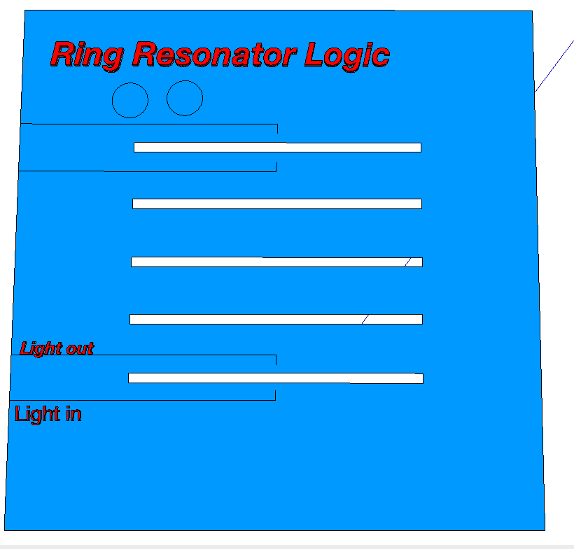
8:00 AM – 8:30 AM Advancing Silicon Photonics for Clinical Applications D. Ratner. Abstract
- This talk was very exciting! The basis for most of his devices’ detection were ring resonators that undergo a characteristic shift in frequency when a sample rests on the surface of the resonator. He uses antibodies and non stick coatings to get specificity. He is able to get picomolar detection in almost real time! Part of the advantage/problem with resonators is that they are very sensitive to environmental factors. One major point of his talk was that we can effectively manufacture enough rings to measure the environmental perturbations and subtract out the shifts. Upwards of 256 devices on a single chip. About 5 years ago, it was impressive to get 4 rings to behave very similarly, with acceptable Q-factor (a measure of the sensitivity of the rings).
- These kinds of technologies are especially compatible with our manufacturing methods, though it would require processing the front end and protecting it before the backside EDP etch. Krishanu has postulated other uses for our membranes as many parallel slot waveguides; I think we could manipulate our membranes as upstream filters and use nanowire waveguides on the front side to specifically illuminate small volumes near the filter surface to track what is coming through the filter.

OP-Sat-2-15.
Glial Cell Engineering / Neural Progenitor Cell and Tissue Engineering
Oral
Saturday, October 25, 2014
1:30 PM – 3:00 PM Room 202A
Moderator 1: Deanna Thompson
Moderator 2: Ryan Gilbert
1:30 PM – 1:45 PM Schwann Cells and Electrical Stimulation: Enhanced Migration and Neurotrophic Factors to Aid PNS Repair L. Zhang, A. Koppes, K. Kearns, D. Thompson. Abstract
- Yet another talk on how cells are targeted to where they are going. There are synergies between neurotrophic factors and electrical stimulation to get Schwann cells to migrate, potentially to sites of injury.
1:45 PM – 2:00 PM Piezoelectric Fibrous Scaffolds for Schwann Cell Induced Spinal Cord Repair Y. Lee, S. Damarju, S. Wu, M. Bunge, T. Arinzeh. Abstract
- Nerve conduit talk. It’s really more about the release of BDNF than the ‘piezoelectic’ part, though potentially spontaneously firing neurons could coax out more BDNF from the scaffold. Some improvement in rats was shown. Can we get the neurons to grow longer/ to specific neurons?
OP-Sat-3-13.
Macro/micro Design for Neurotechnologies / Networked Neural Sensors, Actuators, and Instrumentation
Oral
Saturday, October 25, 2014
3:15 PM – 4:45 PM Room 202A
Moderator 1: Pedro Irazoqui
Moderator 2: Mehmet Kaya
3:30 PM – 3:45 PM System for Integrated Neural Imaging, Recording and Stimulation Z. Liu, H. Cheng. Abstract
- This talk focused on adding EEG information to fMRI to map neural circuits. What’s really cool is that different EEG frequencies that correspond to different modes correlate to different functional circuits!
- Adding the additional information is promising, however questions were raised about the efficient sampling of EEG for deeper cortical circuits (EEG is averaged neural activity, but would be more affected by surface cortical circuits). The time scales involved meant that they had to use low resolution areas to correlate averaged EEG activity to fMRI.
3:45 PM – 4:00 PM Ultrasound Neuromodulation: Field Overview and Observations in the Vagus Nerve of a Rat E. Juan. Abstract
- The abstract really summarizes this talk very well. Not a great presentation.