bEnd3 culture for BBB mimetic (open static system)
Hi all,
Currently I am working on a cell culture system to mimic the blood-brain-barrier (BBB). Essentially, growing endothelial cells (bEnd3) on one side of the membrane, and pericyte and/or astrocyte on the other.
Below is the layout of the cell culture system:
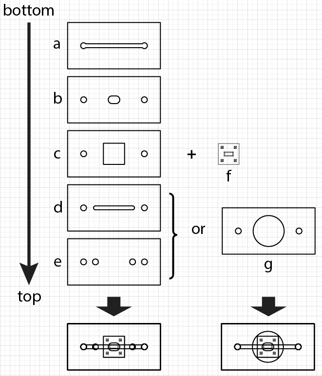
a, b, c, and d are custom-cut silicone gaskets. f is the 5.4mm x 5.4 mm SiN membrane chip (membrane area: 0.7mm x 2mm). e and g are custom-cut plastics (made from the silicone gasket backings) used to guide the cutting and the hole-punching of PDMS slabs, which form the roof of the closed flow system (bottom left) and the well wall of the open static system (bottom right). Adobe Illustrator file of the design (which is drawn to scale) is available upon request (I originally planned to upload it here, but submission of .ai file is not permitted on NRG it seems). NOTE: Gasket “b” is the sealing layer that prevents fluidic exchange between the top and the bottom fluid compartments (so the fluid cross talk can only happen through the membrane). Gasket “c” forms the padding around the SiN membrane chip.
For the pilot BBB run, I seeded bEnd3 in the top well of the open static system. a, b, and c are all 300 μm thick. The bottom-most substrate (below gasket “a”) is a No 1.5 coverslip (2.4cm x 4cm). Pipettes were used to introduce media to the top well (100 μL) and the bottom channel (< 40 μL). The whole system is hosted in a 60mm petri-dish, filled with 5 mL of DI water to reduce evaporation of the cell culture media.
cell culture media: 90% DMEM (Corning 10-017-CV) + 10% FBS + 1% penstrep (final constitution: 100 U penicillin and 100 μg/mL streptomycin).
coating media: 50% cell culture media + 50% FBS. The open static system is UV ozone-treated and autoclaved. The top well and the bottom channel are both incubated with the coating media for either 1 hr or overnight in the incubator.
So the full system looked kinda like this:
![[photo] open static system](https://trace-bmps.org/wp-content/uploads/2015/02/photo-open-static-system.jpg)
============================= DATA ==================================
sample1: substrate incubated with coating media for 1 hr, cell seeding density 15×10^4 cells/mL
day2:
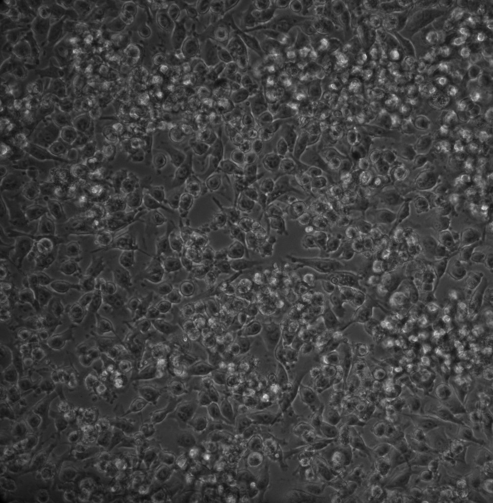 before media refreshment
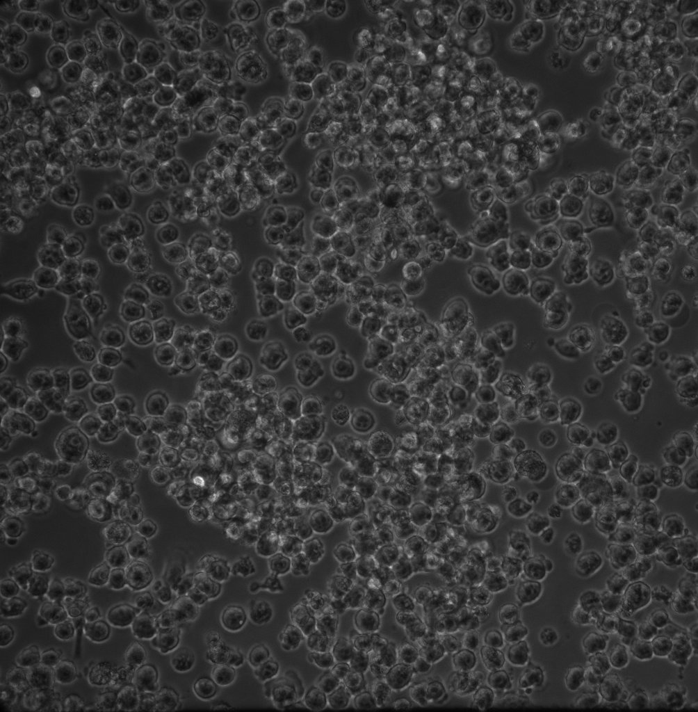 after media refreshment
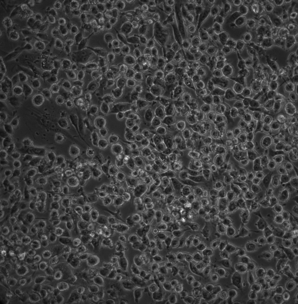
It seems to be the case that many of the spread cells were sheared away during the refreshment of cell culture media (via pipetting). The cell density concentration (15×10^4 cells/mL) also seems to be too high. It is not unexpected since I used 3 times the seeding concentration (5×10^4 cells/mL) that some investigator uses (although, there are others that use even higher cell seeding density, e.g. 10^6 cells/mL). Under the assumptions that longer substrate coating and lower cell seeding concentrations are better for cell adhesion, I prepared “sample 2” accordingly. Assuming that letting the cells sit for 2 days is better for their attachment, I also changed my protocol to refresh my media every other day rather than everyday. (NOTE: For bEnd3 culture in the T25 flask I also refresh the media every other day). I also pipetted slower to reduce shear.
day 4:
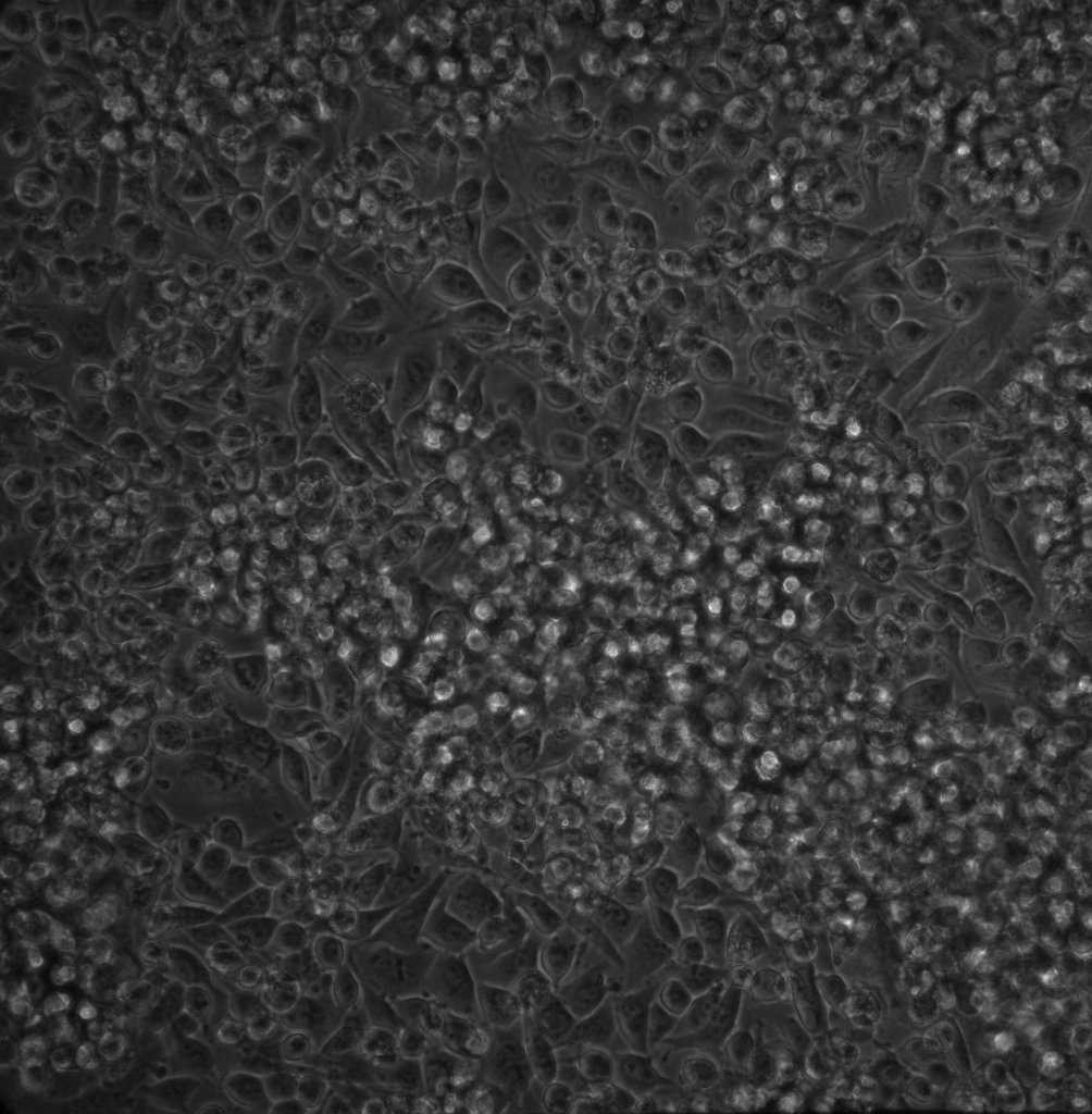 before media refreshment
 after media refreshment
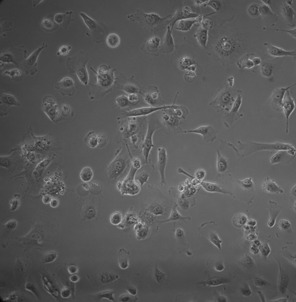
cell detachment still happens, albeit at a lesser degree. Some of the cells that stacked on top of other cells are also sheared away.
day5:
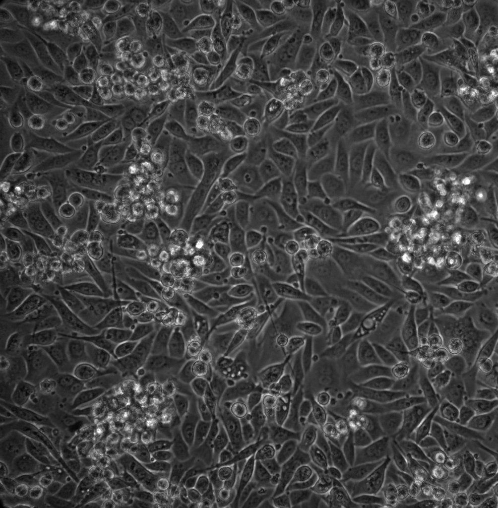

Though some cells are still stacked on top of the others. I suspect these top cells were divided from the bottom cells.
sample2: substrate incubated with coating media overnight, cell seeding density 5×10^4 cells/mL. This sample is prepared a day after sample 1 (from which I learned that detachment could happen).
day 3:
 before media refreshment
 after media refreshment
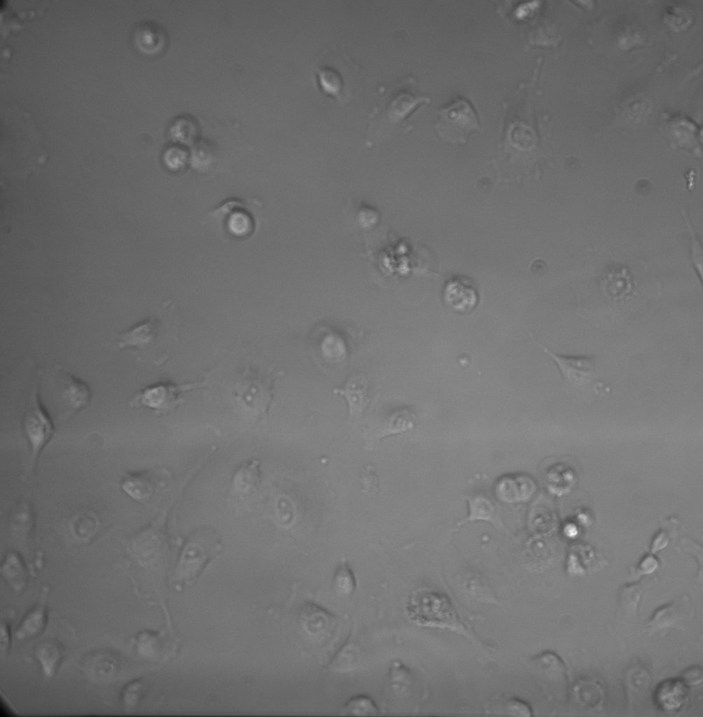
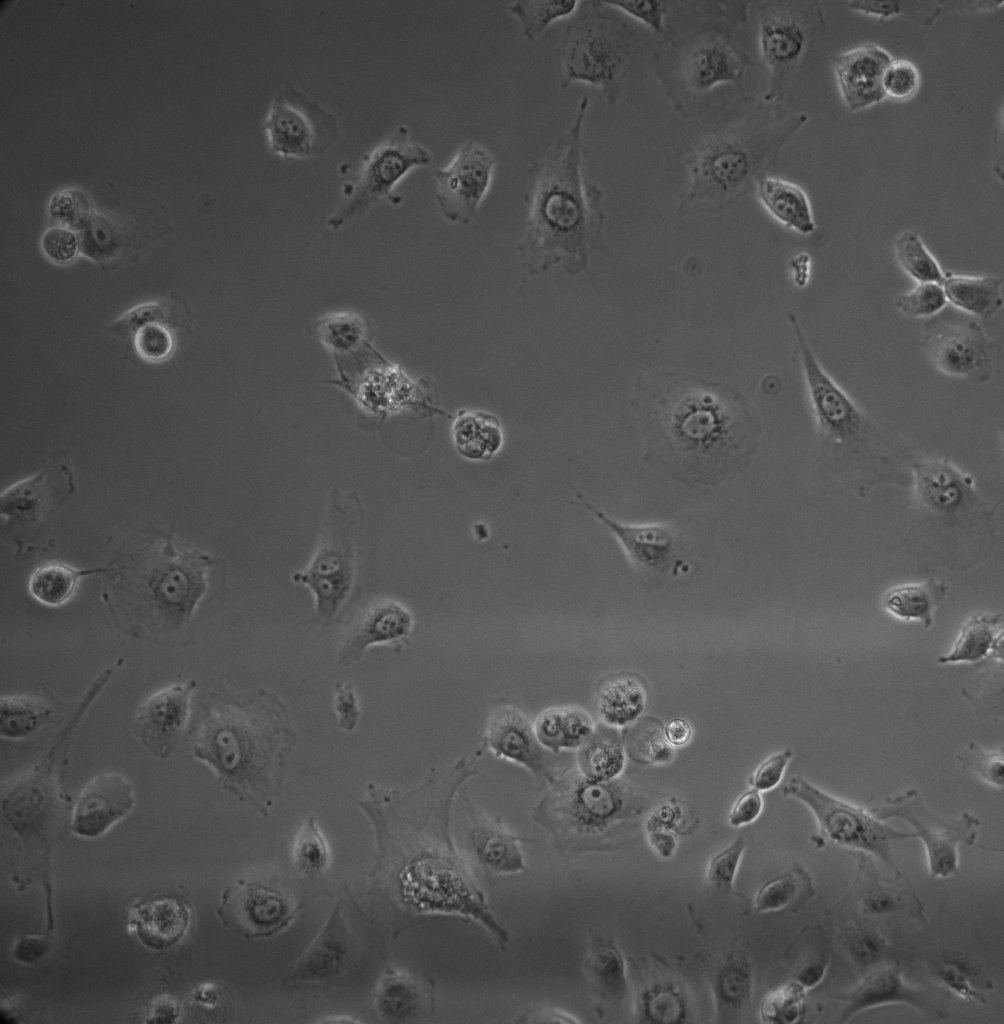
Most cells remained adherent after refreshment of the media. The image before refreshment have worse contrast since somehow there was more than 100 μL on the top well (so the media convexed up).
day4:
Transwell (for comparsion. Cat# 3472, PET (“transparent”) membrane, pore size: 3 μm, area: 0.33cm^2)
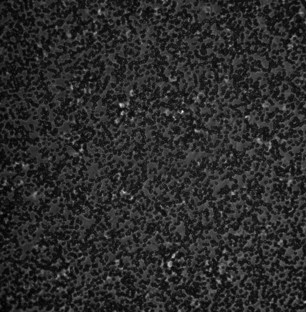 cannot really see much.
Dark circles are probably the 3 μm pores. The white blurs are probably cells.
CONCLUSION: Seems like sample 2 showed better bEnd3 phenotype as far as substrate adherence is concerned. I will stick to this protocol in the future. Maybe I can up the cell seeding concentration to 10^5 cells/mL to speed up confluency. It is also reassuring to see that the bottom channel did not dry out after 2 days, despite its small volume (< 40 μL). And Transwell sucked in terms of bright field imaging.