PDMS Posts
As we were conducting our experiments on dimpled Sylgard substrates we knew that another micro-topography that we wanted to investigate was micro-pillars. In like manner to the dimples, a post microtopography should provide some sort of surface for the cells to “grab onto” and spread as they would on any ECM in vivo. These micro-features impact different cells in different ways that can be inferred from ECM topographies. By analyzing how ADSCs and HUVECs spread and proliferate on these different topographies, we can better understand how they would spread over the ECM in vivo and we can more accurately replicate these conditions in vitro for any in vitro studies. We set out to replicate a technique described “Endothelial cell responses to micropillar substrates of varying dimensions and stiffness” where micropillars were cast from a negative PDMS mold that was constructed from Sylgard 184 that was mixed at a 10-2 ratio opposed to the conventional 10-1 ratio.
First, we took one of our 8um post SU-8 wafers and cast a negative from 10-2 Sylgard 184 as we would to make our substrates. The SU-8 posts should be inspected well and thoroughly cleaned as to ensure that there is no debris which may get translated to the negative mold. After casting the negative (pouring it over the SU-8 posts and allowing it to cure between 70 and 80C overnight), it was treated with acetone which acted as a release agent for the PDMS. After that, they were treated with the corona wand for 2 minutes, followed by a four hour exposure to trichloro-perfluorooctyl-silane in a vacuum desiccator to aid in releasing the positive. From there, 10-1 Sylgard 184 and 10-1 Sylgard 186 were poured over the negative molds to ensure coverage of all of the 2x2mm dimpled windows. The samples were then baked overnight at between 70 and 80C. To release the positive from the negative, the samples were placed in an isopropyl alcohol bath and placed in the sonicator for 180 seconds. Once that was completed, the positive and negative peeled apart relatively easily. This left us with a PDMS surface that had 8um posts on it as well as a reusable PDMS mold for these micropillars.
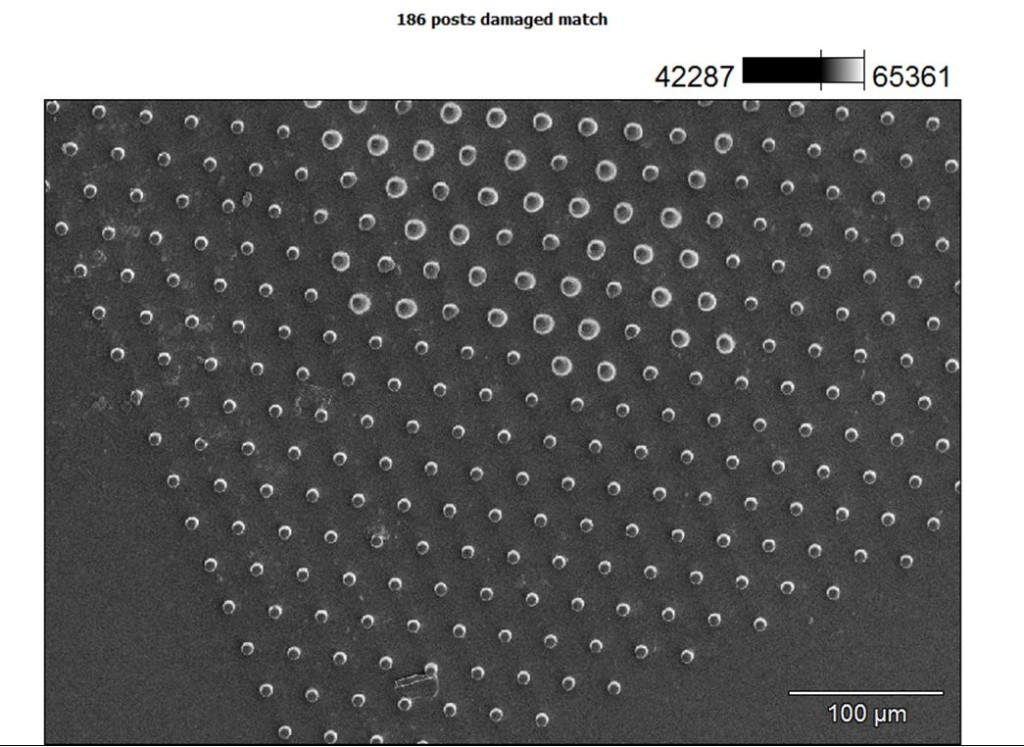
Our results were similar to those in the paper and our only future change right now would be to make sure the wafers are spotless before casting so we don’t distort any dimples in the negative cast which would then be transferred to the positive, micropillared surface. In this image you can see the distortion that occurred in the holes most likely coming from when the negative was cast, we think that it wasn’t cleaned as well as it should’ve been.

Overall, the post liftoff method was successful and subsequent testing showed that we could also reuse the negative molds. That’s good news because now we don’t have to worry about making another wafer to make posts and we can easily make the substrates with simple release procedures and liftoff methods. Also, it gives us another contour to test for our study on cell spreading and proliferation on substrates of varying stiffness and surface topography.