Exosome Isolation Using Tangential Flow and Nanoporous Membranes
Hi all, I’m Kilean. I’m a first year grad student in BME doing my rotations. My rotation project has been working on developing a method that can isolate exosomes from blood plasma. We are trying to compete with techniques such as ultracentrifugation and ExoQuick, which are both the current preferred methods for isolating exosomes.
The ideal method of isolation would be using a Sepcon system, as this is the most user friendly. However, there is a problem with fouling from junk in the plasma and barely any plasma passes through the membrane. Therefore, the task falls upon tangential flow devices and diafiltration to solve the problem.
The chip used in the device came from batch 1156 and has an approximately 29.4 nm pore diameter and a 13.9% porosity. The chip was placed so that the trench side was not exposed to the flow, only the stagnant layer. The bottom channel, which had the stagnant layer, was 2 mm wide. The top channel, which had the flow, was 1 mm wide. Initially, the top channel layer was designed to be 300 μm thick, but this was abandoned in favor of a 100 μm layer. This is to improve the pressure drop across the membrane and decrease the resistance to flow across the membrane, which was important for the first round of trials.
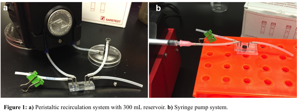
Initially, the idea was to concentrate the exosomes in the retentate, while the proteins and other junk would be collected in the filtrate. Therefore, the thinner top channel was important to achieve significant flow through the membrane. The initial focus was to try and decrease the working volume of the retentate and therefore concentrate the exosomes in the final working volume. In order to accomplish this, the device was connected to a peristaltic pump with a reservoir to recirculate the retentate as shown in Figure 1a. Initially, we started with ultrapure water to test if we could indeed deplete the reservoir and therefore “concentrate” the exosomes. Our syringe pump setup, which was used for later experiments, is shown in Figure 1b.
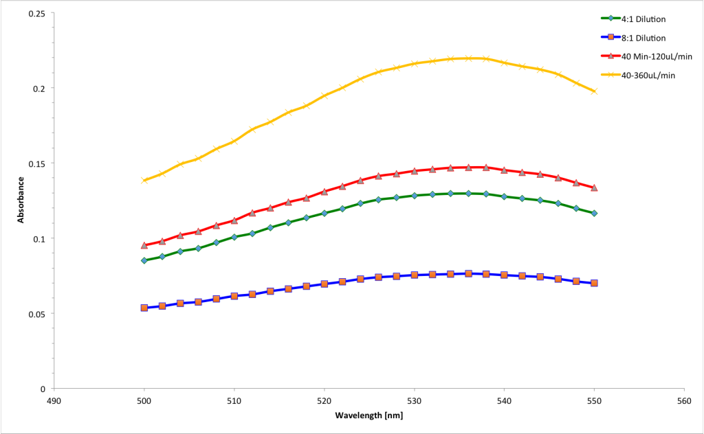
With a starting volume of 400 μL and a flow rate of 120 μL/min, in 40 minutes we were able to collect 198 μL on the permeate side. This means that we increased the initial exosome concentration by a factor of two. This means that for analysis, the exosomes would be twice as concentrated to work with. To put this theory to the test and see if we could achieve the same flow rate with suspended particles, we put a 4:1 dilution of 60 nm gold particles in the ultrapure waster as an exosome simulant. We ran the experiment under the same conditions, but this time only achieved a flow through volume of 69 μL. This was well under half of what we had gotten previously and mildly discouraging. However, using the Tecan, there was a noticeable increase in the absorbance at the gold peak, shown in Figure 2. This suggests that this method would indeed work and provided a promising path forward, despite the necessity for increased recirculation time.
Henry acquired some mouse serum, which we were going to use as a plasma simulant. We would spike the serum with colloidal gold, our exosome simulant, and run this concoction through the device. If there was an increase in absorbance, we would have achieved what we wanted. Also, since we wanted to compete with other methods in terms of speed, we decided to increase the flow rate. We tested the membrane with untreated ultrapure water and found it could easily withstand the maximum pump flow rate. Therefore we decided to risk our systems and run at 1 mL/min.
For the first run, with the gold and serum we achieved a collected volume of a whopping 10 μL in 30 minutes! This was quite puzzling and we though it could be due to fouling, since there was very high concentrations of protein in serum and this could foul the membrane very rapidly. We therefore decided to run pure serum at a lower flow rate to compare and at approximately 500 μL/min we collected around 40 μL of filtrate. We then tested the absorbance of both volumes, which presented another problem. Because of the junk in the serum, the absorbance showed significant background and we therefore couldn’t get a very clear reading on the Tecan to compare results.
At this point, the plasma arrived so we were eager to run experiments with that. Initially, since this was finals week, the aim was to just run it through the membrane and determine if we could see anything that looked like exosomes under the SEM. Therefore, we ran the plasma through the device at 400 μL/min for 30 minutes and rescued the membrane by cutting it out of the PDMS. I should note that several of the membranes were breaking because of the pressure when starting the pump and we probably went through 5-6 devices before actually getting one to work. When we did get the flow through, the extraction was successful but during the drying the membrane broke. We hypothesized that this could be due to the large amount of protein drying on the surface. The samples were imaged, but there was virtually nothing of the membrane to image.
After the break, it was time to revisit the idea of how we would actually isolate the exosomes. It was decided that if there was enough transmembrane pressure, the exosomes would remain on the membrane until that pressure was removed and we therefore wouldn’t have to rely on volume loss concentration. As long as the exosomes were exposed to the membrane, they should be driven to the surface and remain there. We could use a lower flow rate and not have to worry about the low volume that we would collect on the filtrate side. Additionally, there was the problem of drying the membranes so that they remained intact for imaging. Since the top channel was removed, that wasn’t the problem. Therefore, I hypothesized that the bottom channel, which only had a small opening, was the problem with the drying step. There was a small surface for evaporation, which would put a significant amount of stress on the membrane during the drying process. I decided to try removing the back side of the device as well in hopes of retaining an intact membrane. This worked and I finally had a fully intact membrane to image.
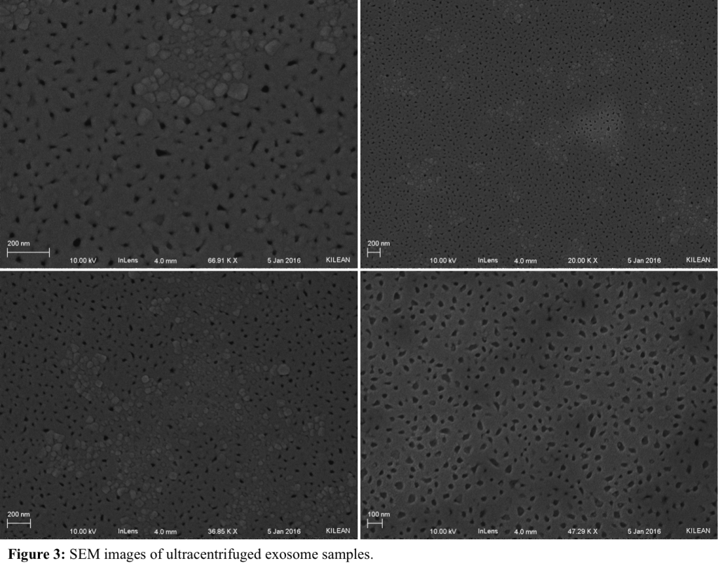
Since we were still unsure if what we were looking at was exosomes, we decided to perform ultracentrifugation to isolate the exosomes. I followed a protocol found in JOVE [1] with a modification of a 90-minute, 100,000 x g run time in the ultra centrifuge. Jamie had mentioned performing a prefiltration step on the plasma using a 0.2 or 0.7 μm filter to remove unwanted junk. We tried this with both sized filters and they immediately fouled. There was a lot of protein in the plasma and this concerned us for a run through the device. We therefore decided to skip the prefiltration and went straight to the ultracentrifugation. The pellet, which was not really visible, was resuspended in 100 μL of PBS. As we had been breaking several membranes using the peristaltic pump, we decided to switch to a syringe and do only one pass through the device. The SEM images of the results are shown in Figure 3. There were particles blocking the pores, which are thought to be salt crystals from the PBS, but we couldn’t get the EDX to confirm this. We could therefore not conclude that we could positively identify exosomes and turned to someone with experience in the matter for advice.
Ryan Dawes in Ed Brown’s lab is working with exosomes for cancer and we decided to talk to him to get advice on the ultracentrifugation protocol and any other pointers. When we went over there to talk to him however, it turned out that he used the commercially available ExoQuick to isolate his exosomes. He had incredible success and showed us some TEM images of their exosomes. Their NanoSight counter put the concentration of exosomes in mouse plasma at something x 1011 per mL of plasma. This was significantly higher than our previously accepted value of 105-106/mL. We attributed this to the ExoQuick prep and decided to try this method to establish a baseline. However, Ryan had never worked with frozen plasma and warned us of literature that pointed to a decreased amount of exosomes in frozen plasma. Therefore, Henry graciously donated fresh blood for our experiment and we would compare this to the frozen plasma, as well as to plasma untreated with ExoQuick.
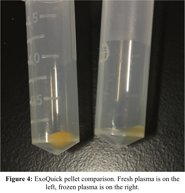
Immediately in the isolation, we noticed a significantly smaller pellet in the frozen plasma sample, as shown in Figure 4. The fresh plasma had a very noticeable pellet, which gave us high hopes. These pellets were resuspended in 300 μL of PBS. The syringe pump was set to a 10 μL/min flow rate, which would give us an experiment time of approximately 13 minutes. This allowed us to pass about 100 μL though the device while accounting for dead volume in the syringe. We had been using ultrapure water as our filtrate liquid, but decided to switch to PBS in order to minimize lysing. We ran the two ExoQuick samples in duplicate and one of each fresh plasma and frozen plasma. We also had control samples to just characterize exosomes, but have yet to get around to imaging these samples with limited SEM availability.
The SEM of the fresh plasma treated with ExoQuick showed that there were particles on the membrane with smooth, rounded edges on the membrane, but these were concentrated to the flow inlet of the membrane. A series of pictures taken along the length of the membrane are shown in Figure 5. There is obviously something fouling the pores on the membrane and could possibly be remnants of the ExoQuick polymer. The frozen plasma treated with ExoQuick showed similar characteristics and these images are given in Figure 6. There was a distinct leopard-print pattern on one of the samples, which was probably an artifact of drying. The particles on this sample tended to be concentrated in the lighter areas, while the dark areas had clean pores.
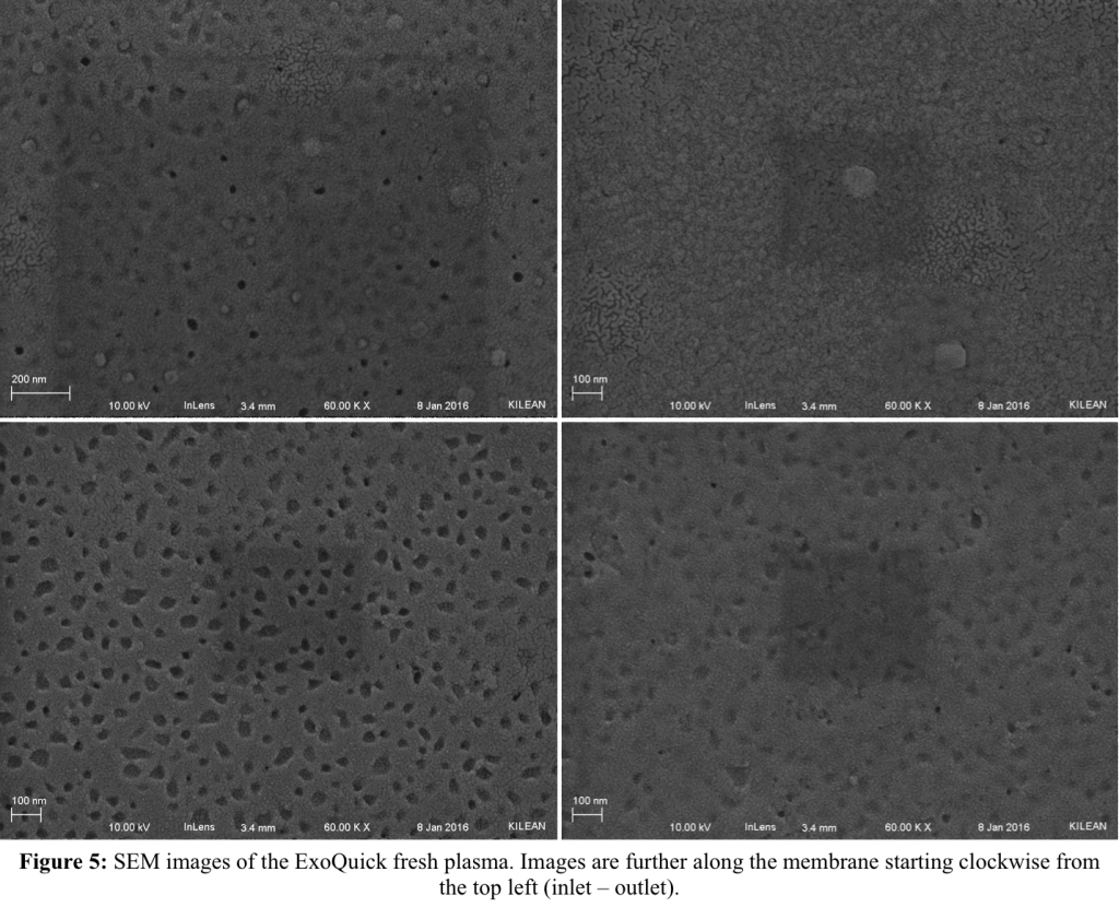
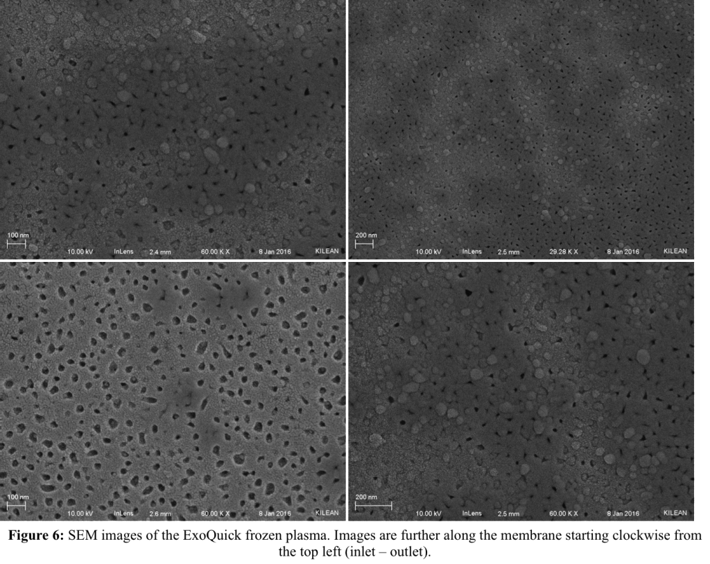
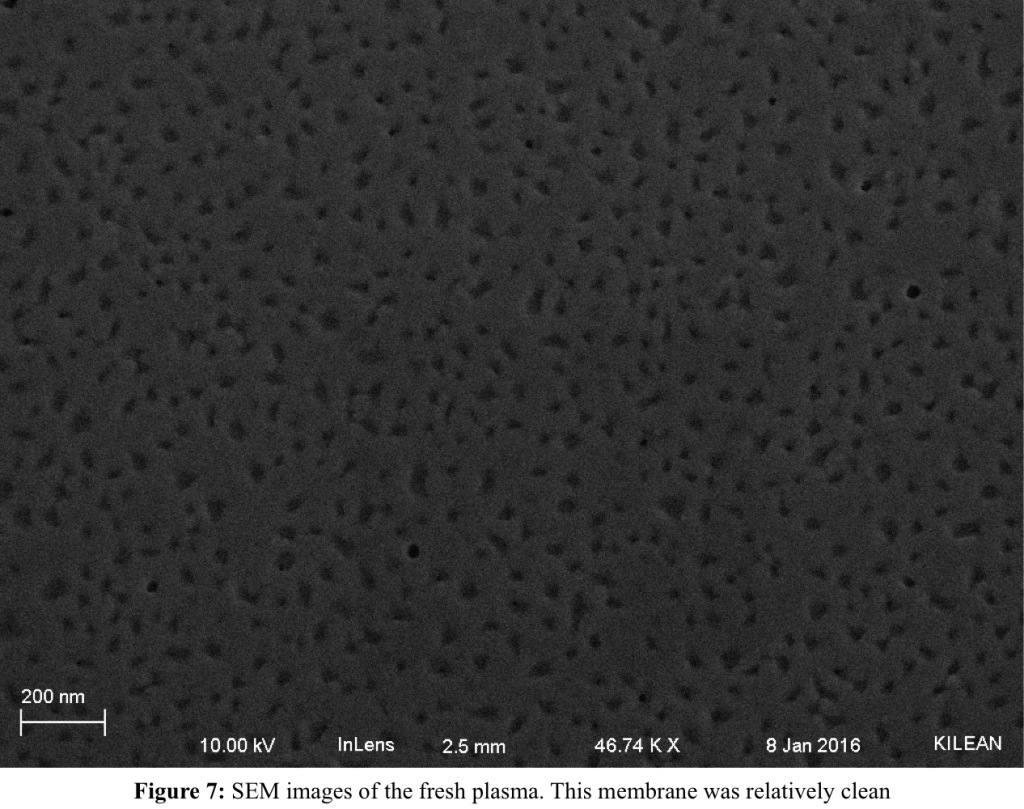
Figure 7 shows the fresh, untreated plasma membrane. It should be noted that while the membranes that had exosomes isolated with ExoQuick were intact and survived the drying process, the membranes with pure plasma both broke and there was very little membrane to actually image. Indeed, for the frozen, untreated plasma there was not even any membrane left to the naked eye. However, this did not stop us from imaging it and Figure 8 shows the results from that sample.
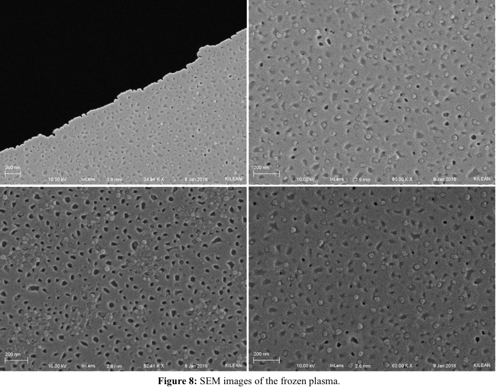
It would appear that through the seeming loss of the sample, there were some very significant results, as the spherical objects inhabiting the pore areas would appear to be exosomes. They were all over the surface of the fraction of the membrane that was left and would suggest a successful experiment. If the straight up plasma produces the result of exosomes all over the surface, then there is no need for the ExoQuick prep as there is an exosome in nearly every pore. We ran energy dispersive x-ray spectroscopy (EDX) on the samples to determine if the particles were salts and there was no signal aside from background for sodium, phosphorous and chlorine. Figure 9 shows the spectrum, along with a comparative spectrum from a salt crystal on one of the other membranes.
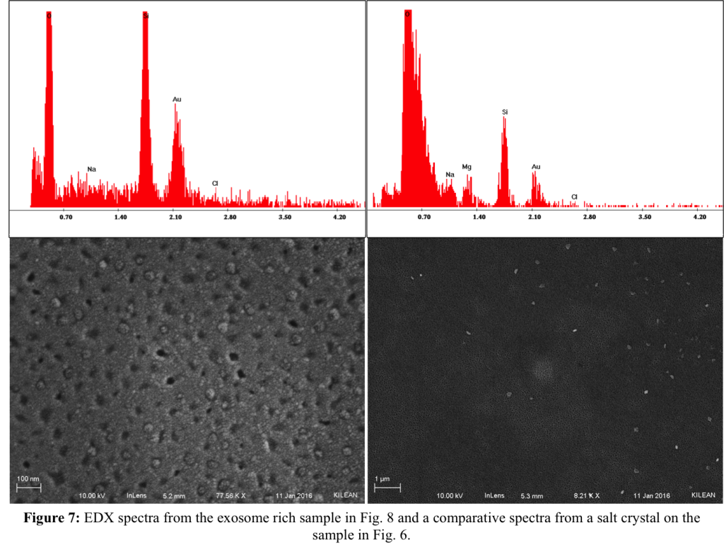
There is need for future work for this data to be conclusive. As this is only a fraction of one sample, we need to (and will) run more samples to confirm repeatability, this would also appear to suggest minimal protein fouling with the straight plasma, which is an ideal result. If these results are indeed repeatable, we could then move forward with other tasks such as definitively identifying the particles as exosomes, ensuring that the exosomes are mostly caught on the membrane and are not all being lost in the flow through or when we are drying.
Sorry for the long post, but hopefully it is informative!
[1] Lässer, C., Eldh, M., Lötvall, J. Isolation and Characterization of RNA-Containing Exosomes. J. Vis. Exp. (59), e3037, doi:10.3791/3037 (2012).