Ultrathin Polymer Membranes with patterned, micrometric pores for organs-on-chip
Paper for Friday’s Journal Club … Watch this space for a summary and analysis.
Synopsis
- 2um(?) pores in 100 nm thick PLLA. Very, very low porosity.
- Fabricated with a PVA microneedle mold produced with femto second laser.
- Microfluidic device integration
- Claims about cell (HUVEC) viability and growth are poorly supported. Cells are essentially on non-porous PLLA.
Abstract
- These people need to learn about silicon nanomembranes.
- The claim that submicron thickness is essential for paracrine signalling should be supported – best evidence found in?
- Existing methods are expensive, time consuming, yada yada.
Methods
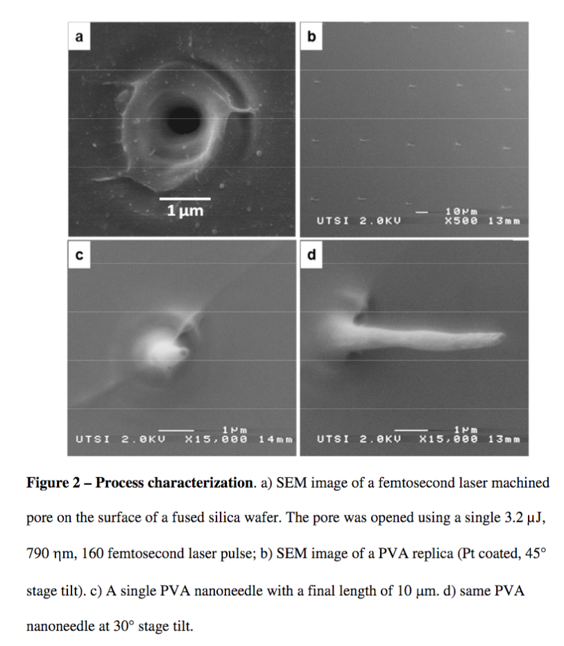
- Two molds. Start with femptosecond laser puled pores in 500 um fused silica wafer (glass). Mold 1: (201)^2 pores -> 40,401 pores per cm^2 = 127,000 um^2 / cm^2 = 0.127 % porosity, P-P spacing of 53 microns. One 2 um pore per cell, maybe. – Note that they never discuss this obvious number. Mold 2: 16 x 100 pattern on 6 mm x 1 cm so 2,667 pores per cm^2 = 0.008% porous with 100+ um between pores.
- 10 um deep 200 um indents with laser then KOH etch.
- PVA in alcohol painted on with a brush, dried, and removed. Apparently fills pores to create microneedles.
- Spin coating PLLA on PVA. Any verification of thickness? Profilometer or SEM cross section.
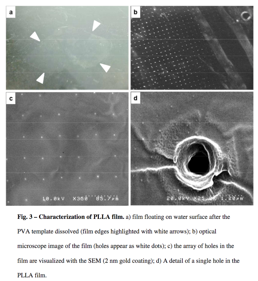
- Transfer to PDMS done through contact and PVA dissolution in deionized water.
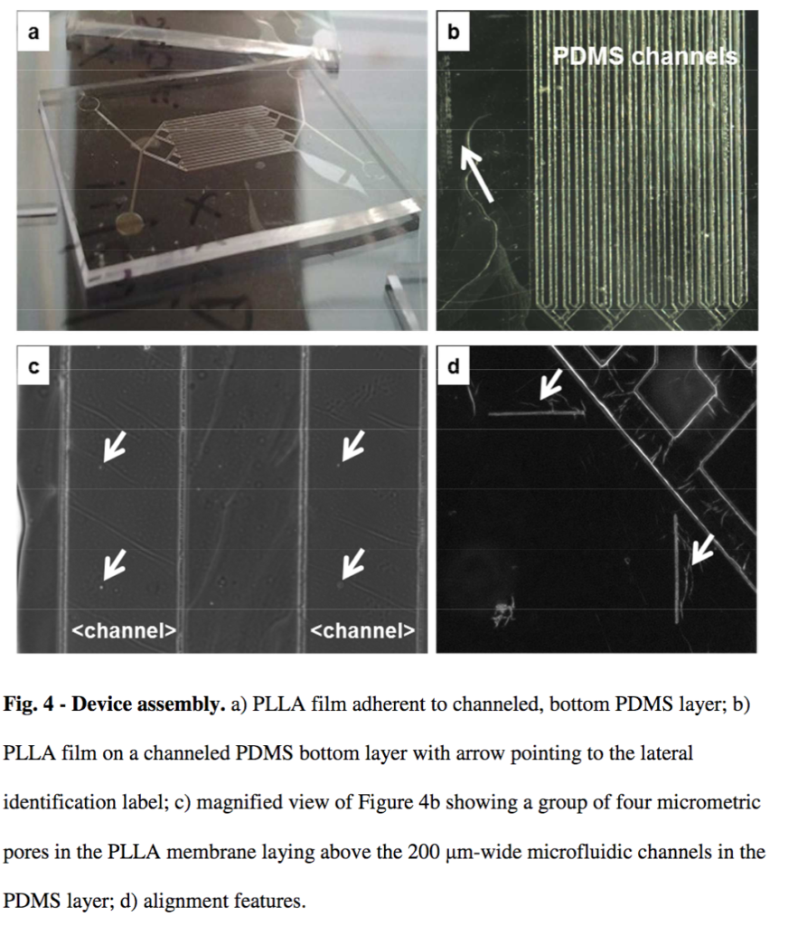
- Columns of pores aligned with 16 microchannels
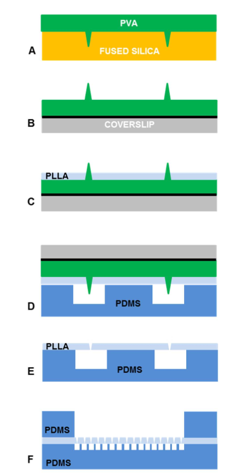
Fig. 1 – Process for fabrication of a semipermeable ultrathin PLLA membrane. a) PVA microneedle array is created by casting the PVA sol
ution on a fused-silica mold and by allowing the PVA to dry. b) PVA replica is lift-off bonded to coverslip using adhesive tape (black solid line). c) PVA replica is spin coated with PLLA. d) PLLA film is placed in contact and aligned to the PDMS microfluidic layer. e) PLLA film is adherent on the PDMS layer. The final assembled device presents the PLLA barrier between two microfluidic chambers.
 Results
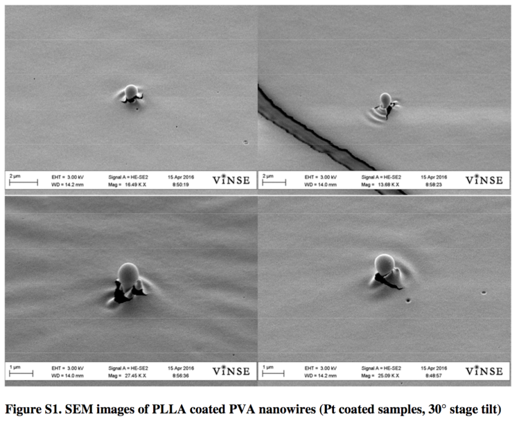
- 1 um pore size here, 2 um in the intro? Can’t claim uniformity without showing multiple measurements. Why 1 um pores?
-
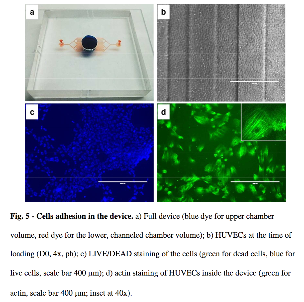
Cannot make any claims about growth rates from figure 5C-D? If anything a lack of confluence at 7 days suggests growth is slow. Show control morphology.
-
Viability – must quantify and compare to controls. One control should be nonporous PLLA (but that is essentially what this is).
-
Transparency would suffer if pores were more numerous. Claim is meaningless.
Discussion
-
“With the current system, this translates to patterning a regular array of four million surface pores inside a one square centimeter area in less than one hour. Molds can be prepared with a distance between pores as small as 2 μm.” – They should do this. Make a very high porosity membrane. They are currently not even close.
- The authors hint here and in the intro that this technology might be used to control molecular passage. Language needs to be more careful. Microporous material cannot do that.
Finally – what the hell is this?