NSL fabrication – Released membrane pictures
This post includes some optical and SEM micrographs of a SiO2 membrane patterned by nansosphere lithography using 300 nm polystyrene beads. An SU-8 microgrid was deposited on top and the membrane was released by dissolving a bottom layer of ZnO. After the release, the membrane is rinsed in water and transferred to a clean substrate. The first thing I noticed was the time needed to release the membrane, which significantly reduced (from days to a couple of hours), this was strong indication of the presence of throughpores. Below are some pictures taken with an optical microscope from a released membrane, resting on a Si wafer.

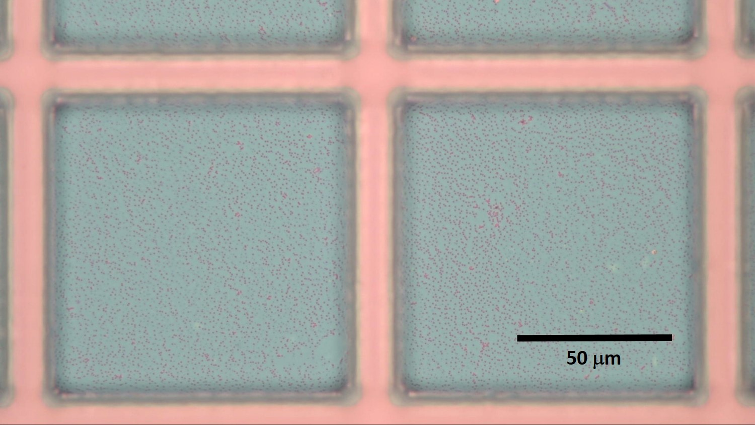
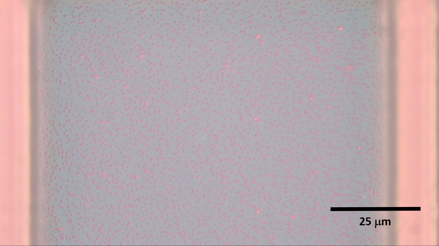
From these pictures you can see there are some pore doublets, triplets, which form from beads that are too close. Not all the pores exhibit clear edges which might be due to the microscope not able to resolve those “smaller pores” or they may not be throughpores.
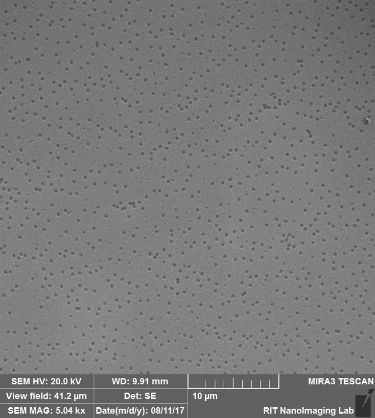
The same membrane was imaged in the SEM (thanks to Aslan), the sample was coated with Au for 40 secs. The following two pictures were taken from the top side of the membrane. Doublets can be observed as well as half pores (smaller features that could correlate with the feature the OM is not able to resolve) and an agglomerate of beads which resulted in pores connected through “channels”.
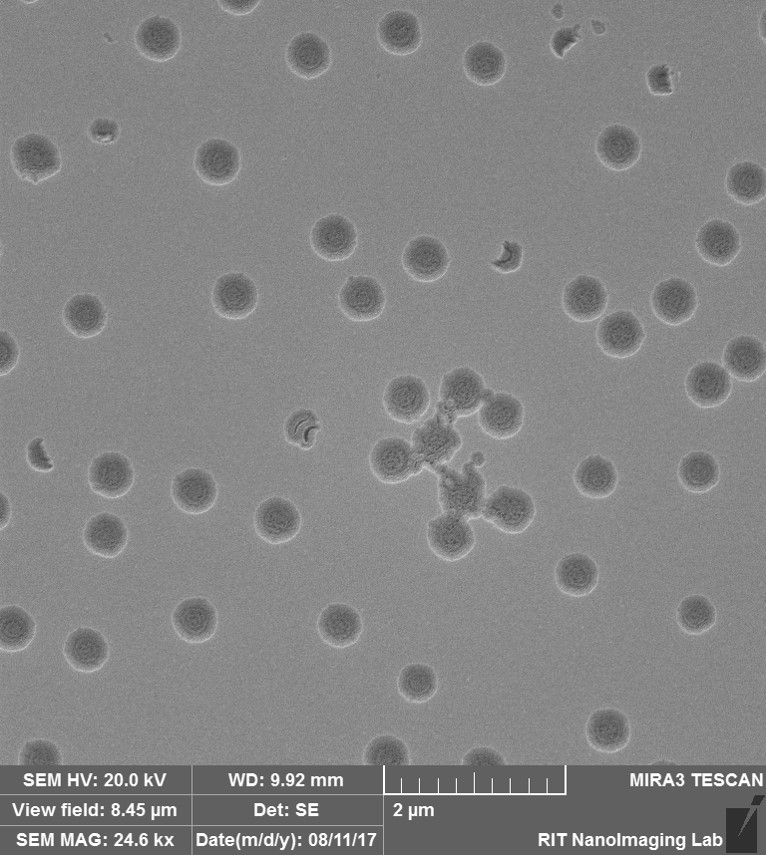
Here is an image taken at higher magnification.
The following two pictures were taken from the bottom-side of the membrane, from a corner where the membrane was curled. (More defects can be seen here, remember this is a corner so higher and uglier defects are expected).

Here is the same area at higher maginification.

We will integrate this membrane into a simple device to test dye permeability to evaluate the presence of through pores.