Latest Update on Wound Healing Study
Introduction:
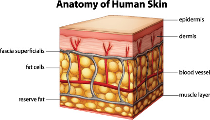
Fibroblasts: Dermal fibroblasts are cells within the dermis layer of skin which are responsible for generating connective tissue and allowing the skin to recover from injury. A fibroblast is a type of biological cell that synthesizes the extracellular matrix and collagen, produces the structural framework (stroma) for animal tissues, and plays a critical role in wound healing. Fibroblasts are involved in key processes such as breaking down the fibrin clot, creating new extra cellular matrix (ECM) and collagen structures to support the other cells associated with effective wound healing, as well as contracting the wound.
ADSCs: ADSCs are histologically located beneath the dermal fibroblasts in the skin. Thus, growth factors secreted by ADSCs may diffuse into the dermis and epidermis in cases of skin damage and accelerate wound healing. ADSCs are known to induce angiogenesis and promote healing in animal models of diabetic skin wound mainly through paracrine secretion of angiogenic and antiapoptotic factors, such as vascular endothelial growth factor (VEGF) and fibroblast growth factor (FGF2). In vitro studies have demonstrated that conditioned medium (CM) prepared by ADSC culture (ADSC-CM), which contains angiogenic growth factors such as VEGF, basic FGF2 and hepatocyte growth factor (HGF) enhanced type I collagen secretion and migration of cultured human dermal fibroblasts, and stimulated proliferation and migration of cultured human keratinocytes. One of the other factors that ADSCs secret is exosomes (30 – 150 nm). We decided to investigate the effect of exosomes derived from ADSCs on Fibroblasts migration in wound recover.
Method:
One of the most popular cell migration assays is the scratch wound assay, which is inexpensive, simple to implement, and can be run with several samples in parallel. Typically, cells are seeded into a multi-well plate and allowed to proliferate to confluent monolayers. A portion of the confluent monolayer is then scratched off, typically with a pipette tip, to create an artificial wound. The rate of wound healing can be determined by the reduction of the wound size over time. In most studies, the wound size is reported based on area or by the distance spanning between the opposing wound edges.
Ascione et al. showed that the report of wound healing by area tend to exhibit high variability even amongst the same sample types. Furthermore, it is difficult to assign a definitive boundary to represent the wound leading variability that arises during the user arbitration of the wound edges. In addition, the variation in cell density and wound size seem to be ignored in literatures. These two factors have important effect on the final result of area recovery.


Also, defining where the wound is recovered is challenging by area recovery. In most of the papers, authors have considered the wound closed or recovered as soon as the left and right side leading cells contacted each other without considering the gaps still remained between the cells.
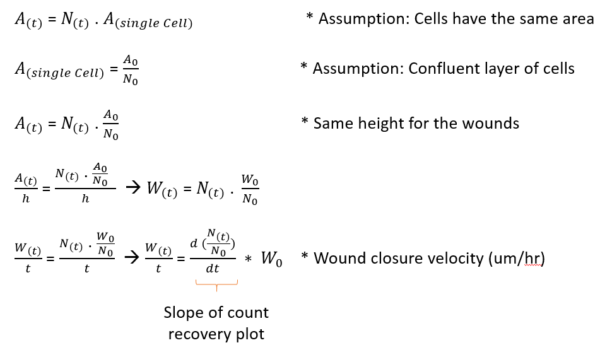
Conceptually, a wound can be considered fully recovered when the same number of cells before scratching move to the wound after scratching. An alternative approach to area recovery is to monitor the number of cells in the wound space over time which is called count recovery. Count recovery can be defined as the ratio of the number of cells migrating to the wound region over time to the number of cells in the wound before scratching (Nh/N0)). The count recovery is independent of the cell density but it still depends on the wound size.
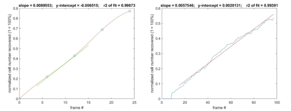


Therefore, a new factor is needed in order to be able to compare samples with different wound size. We are proposing “ Wound Closure Velocity” as the studying factor in wound healing studies over the recovery. The “Wound Closure Velocity” can be calculated using the count recovery.
Count recovery plot over time consists of a linear region which is the recovery phase and then the plateau which is coming from the remodeling phase (This can be seen in the video). The slope of the linear region is an important factor called “Count Recovery Rate”, even though this factor still depends on the wound area.
Literature review:
Most of the literatures studying the effect of exosomes derived from stem cell on the migration and the wound recovery have been done by isolating exosomes from cell culture conditioned media by different techniques such as ultracentrifuge and Exo-Quick and then adding them to fibroblasts. Although, it is important to note that exosomes have been isolated from a high volume of conditioned media resulting in high concentration of exosomes at the end. High concentration of exosomes derived from stem cells have been shown to help the recovery of the wound compared to the control. But the real question is, how about in a physiological ratio of ADSCs and FBs? (Lower concentration of exosomes).
In this project, we came up with a direct co-culture system for studying the effect of ADSC-Exo on fibroblasts recovery. But the question is what exosome protein concentration this system has compared to the used concentration in literatures?
According to this paper published in 2017;
They cultured 5 flasks of MSCs in T25 (25 cm2) and when they reached 80 % confluency they changed the media with exosome free media for one more day and then they collected the conditioned media and isolated the exosomes from that. They reported that this 125 cm2 surface area of cells had 100-200 ug of exosome proteins. In our system, we have 0.9 cm2 of surface area covered by ADSCs which gives us ~ 1ug of exosome proteins after 1 day of the experiment.
We designed two sets of experiments; first studying the effect of the media (Complete media, Exosomes free media, and Serum free media) on wound closure velocity and second studying the effect of different combinations of cells (ADSC-FB, FB-FB, and 0-FB) on wound closure velocity.

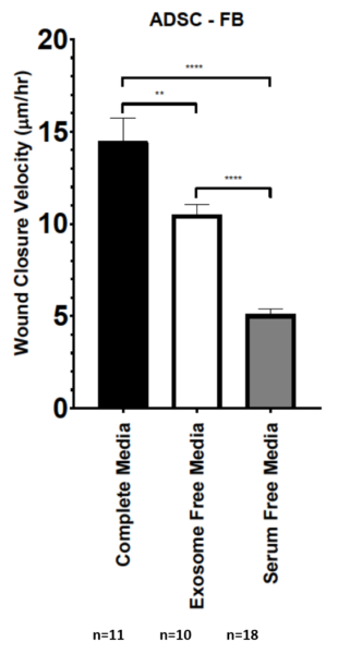
- Different Media Effect:
ADSCs were cultured in the outside region and FBs were seeded in the inside region and after reaching 80% confluency, the FBs were scratched using 20-200 uL pippete tip. Two different experiments (24 well plate ) were performed to compare the effect of complete media, exosomes free media and serum free media. Serum free condition is basically the background condition. Assuming that during the process of depleting exosomes from complete media, only exosomes were depleted and all the other nutrition were still in the media, the difference between exosome free media and serum free media is related to the non-exosomal nutrition in the FBS and the difference between complete media and exosome free media shows the effect of serum exosomes on the wound closure velocity. Comparing these 2 values shows that non-exosomal nutrition in FBS have slightly higher impact on the wound closure velocity compared to serum exosomes in the ADSC-FB system. (Maybe we should concentrate these for wound healing instead of exosomes?)
2. Different combinations of cells:
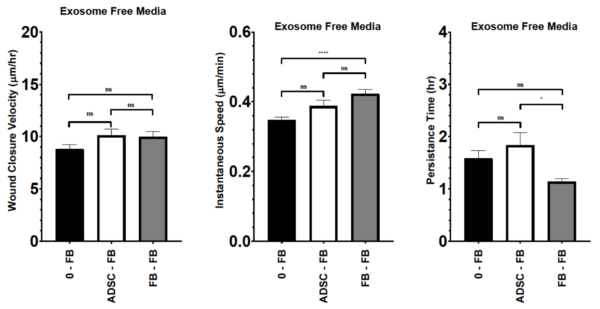
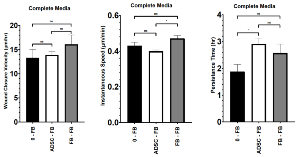
3 different combination of cells were designed including (ADSC-FB), (FB-FB) and (0-FB) and the wound closure velocity of Bfs inside was studied under 3 different conditions ( serum free media, exosome free media, and complete media). No significant difference was observed between different combination of cells, suggesting that ADSCs in this ratio of cells to FBs are not helping the recovery under any of the media conditions.
Consistent with our first set of experiment, for all cell combinations, same trend for wound closure velocity was observed with the order of complete media, exosome free media and serum free media.

More detailed information of each data set:
We were also interested to study the instantaneous speed and persistence time which technically are 2 factors important for wound closure velocity. For serum free media, No significant difference in the instantaneous speed and persistence time was observed between the conditions. For Exosome free media, FB-FB condition showed higher instantaneous speed compared to the 0-FB but lower persistence time compared to the ADSC-FB. For complete media, FB-FB showed higher instantaneous speed compared to ADSC-FB and ADCS-FB had a higher persistence time compared to 0-FB.

It is important to note that these results are coming from different experiments, for future experiments, i would like to repeat some of these experiments. Also we are planning to repeat the different media with 0-FB and FB-FB.