Barcikowski Collaboration Update
Over the last week or so I’ve taken some lovely SEM pictures with Joe, used the light scatter, and ran some separations on filters I assembled myself. Someone from the Barcikowski lab, Dr. Rehbock, got in touch with me and sent me an updated description of the four samples they’re sending and what they want done with them:
1) 7-10 nm bioconjugated gold colloid with peptide ligands in the supernatant (pH=9); retentate free of ligands
2) Bimodal gold colloids with fractions 7-10 nm and about 20 nm (pH=9); permeate with fraction < 10 nm
3) Monomodal gold about 10 nm (pH 6-7); permeate < 10 nm
4) Polydisperse platinum NPs (4-40 nm) (pH 6-7); permeate with fraction < 5 nm
I plan to use next week to do some similar separations, ie separating out 5nm gold out of a mix of 5 and 10nm stock, so that when the samples come I can just troubleshoot instead of starting protocols from scratch. The Samples are due to arrive by the end of April.
After assembling some 20nm filters from scratch I used them to separate 5nm, 10nm, 15nm, and 20nm gold. I used the standard curves from my previous experiment to get the percentages.
For the 5nm gold particles, the average dilution was 33% (where 100% was the 1:1 dilution of the stock solution in your drawer)
for 10nm – 41%
15nm – 4%
20nm – 3%
Note that 10% is the cutoff for the machine, so that both the 15nm and 20nm results are noise.
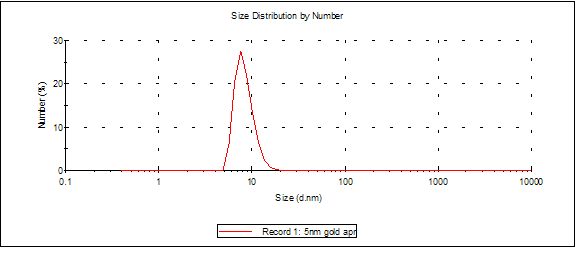
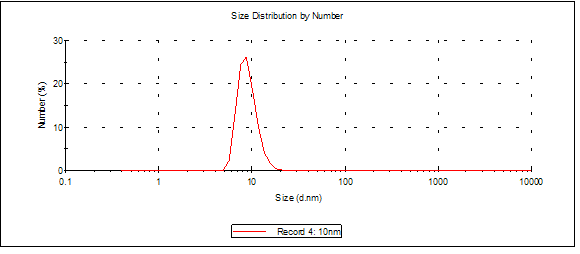
I used the light scatterer on the filtrate of my separations. The results were as follows:
5nm gold had its largest number PSD peak (averaged over three samples) at 9.09nm:
10nm at 8.86nm:
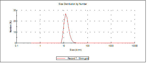
15nm at 13.2nm:
20nm at 22.6nm: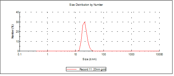
I think this means that the 5nm samples are clumping up preferentially in pairs, but everything else looks fair.
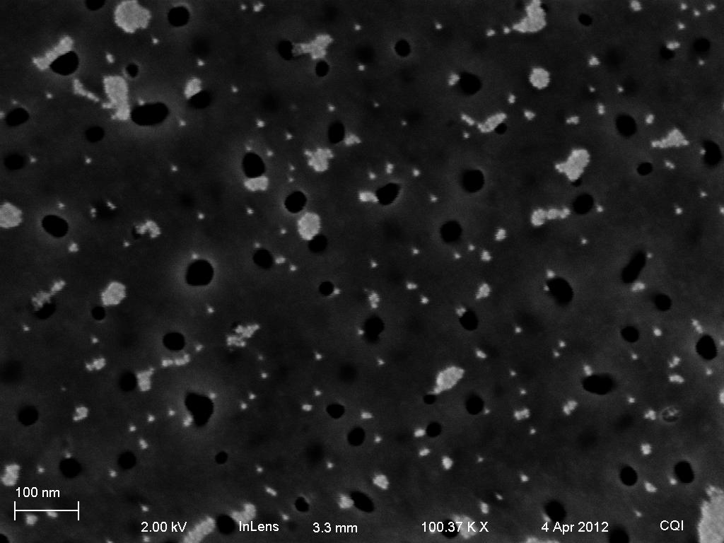
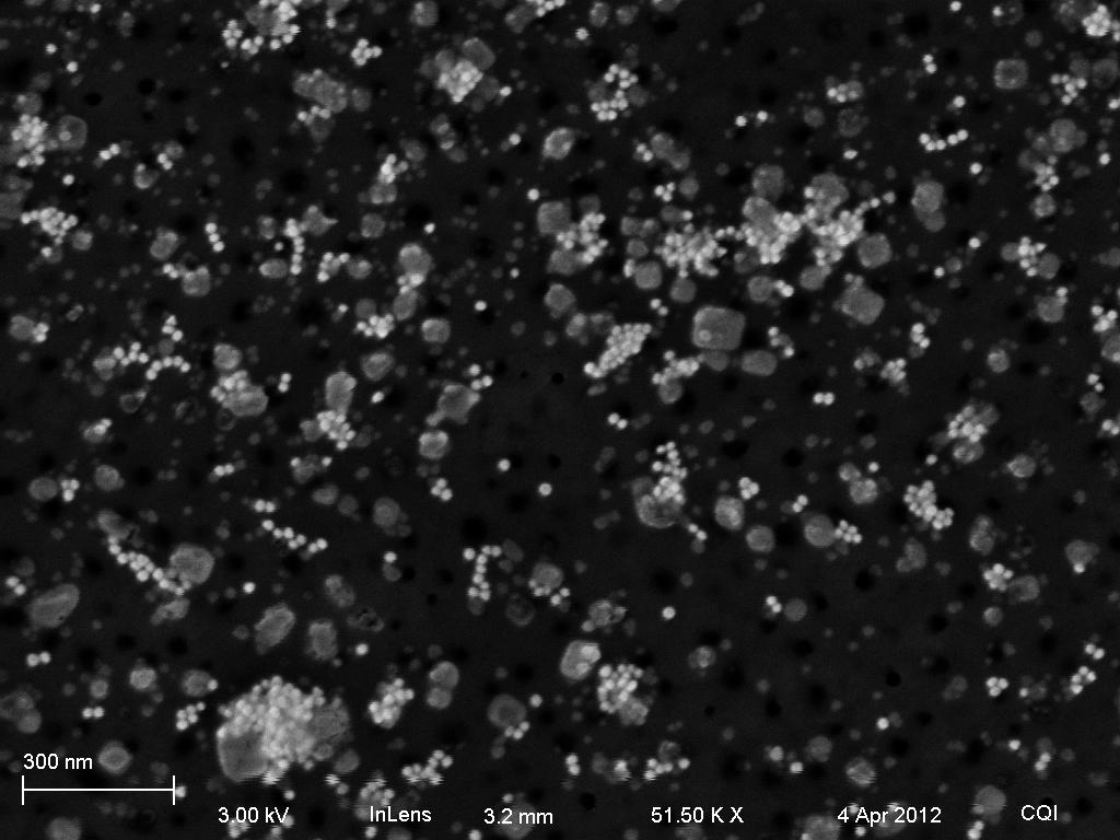
Four representative pictures of my SEM session with Joe are below: