CMFDA Dye Potentially Harms Cells
Invitrogen offers a number of stains and dyes for use in tracking cells and fluorescent imaging of tissue culture. One prevalent live stain is called CMFDA (5-Chloromethylfluorescein Diacetate), a green fluorescent molecular probe. This stain is utilized for tracking cells and monitoring growth & viability of the cells.
While performing studies using CMFDA, there appears to be negative effects on the cell lines.
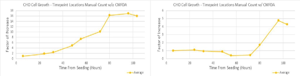
When looking at CHO cells which had uptaken the CMFDA and were exposed to excitation light, there appeared to be a delay in growth that lasted roughly 24 hours. Due to the robustness of the line, they renewed propagating after this and followed a fairly normal growth curve.
The bEnd.3 cell line was impacted more heavily. Those cells which uptook the CMFDA and were excited with the fluorescent light within the first day started rounding up, did not propagate, and either died or migrated to other areas (though it appears that cell death is likely). Based on these observations, the use of the CMFDA dye appears to be potentially toxic to cells to varying degrees, depending on the cell line.
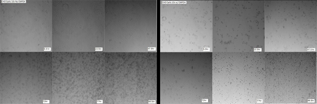
The lab purchased CMFDA to be used in characterization of cell proliferation and growth, and to model growth curves of the cell lines used in the lab (previously CHO [Chinese Hamster Ovary] and bEnd.3 [mouse brain endothelial], but now only bEnd.3 are being focused on with the CMFDA). Invitrogen states that normal concentrations of CMFDA to be used (diluted first into 10mM concentration in DMSO) is between 0.5 uM -> 25uM (second dilution in serum-free media). For the experiments in this post, either 5uM or 7.5uM concentration was utilized. Hams F12 media was used with CHO cells (below, left), and DMEM with bEnd.3 cells (below, right).
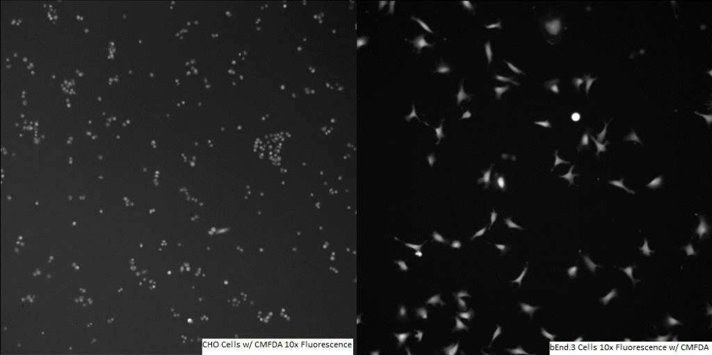
Whilst working with the CMFDA stain initially, it seemed to function as expected. The stain fluoresced well, and identification of the individual cells was more or less straightforward. Counts did not produce normal growth curves however, and there were some other issues that began to arise.
The first problem to become apparent was the loss of fluorescence by spread CHO cells. In images after roughly 48 hours (but to a lesser extent also earlier, even less than a day after seeding and staining), fluorescence was still visible but only on more rounded and “pseudo-adhered” cells, not on those which were more spread (which normally indicates potentially happier cells).
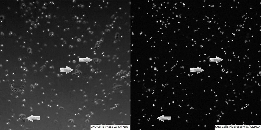
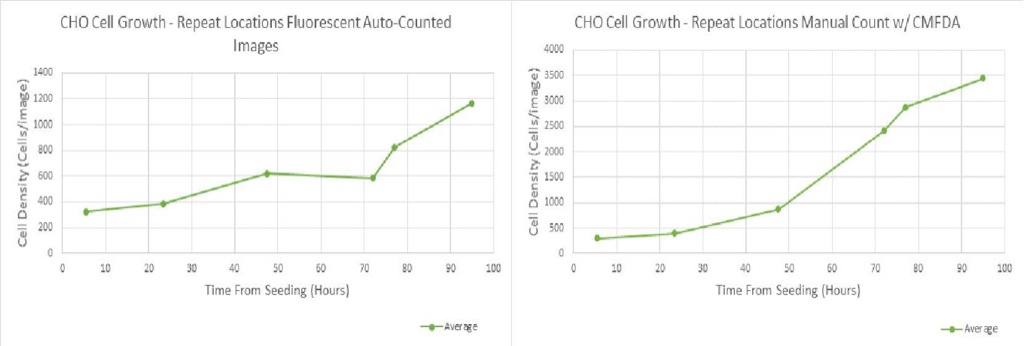
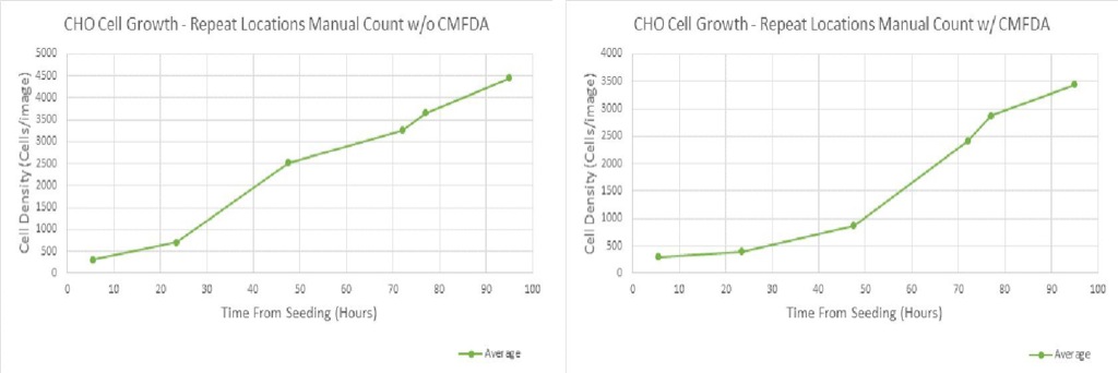
This made counting via the fluorescent images inaccurate and detrimental to growth curves, as they no longer represented the actual growth that occurred. Manually counting the phase images of the same location and time showed that the counts based on the fluorescent images were off by up to >2000 cells (~66% of total number of cells in image, see comparative charts).
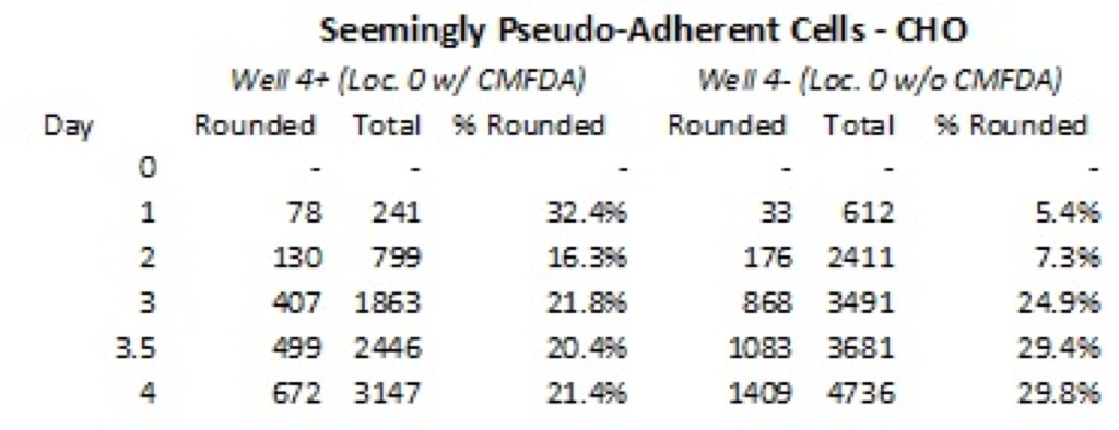
In addition to the problems with counting, it appeared that more cells were rounded up by percentage in locations with CMFDA present. These percentages were determined through counting of roughly equivalent wells. The apparent jump in the well without CMFDA to almost 25% from just over 7% is caused by the development of a confluent monolayer (and subsequently growing cells would have a more rounded appearance and be less adhered). With CMFDA, the cells have a consistently higher (>15%) rate of roundedness. The first timepoint was not counted, due to the fact that the cells were still settling and therefore looked predominately rounded in both wells.
On the final of the CHO trials, it appeared that an approximately 24-hour delay in cell growth was caused by the CMFDA. In those wells that had the stain, very little growth occurred in the first day, though after this it seemed to normalize to what would be expected for the CHO cell growth curve based on non-stained cells. Both observation of the images and the growth curve counts supported that this was the case, though the reason as to why this happened is presently not fully clear.
After finishing with the CHO trials, I moved on to using the bEnd.3 cell line. A coworker had previously dealt with using CMFDA and bEnd.3 cells, and hadn’t reported anything untoward. Additionally, it appeared that spread cells still fluoresced well and there weren’t any issues with miscounts for that reason.
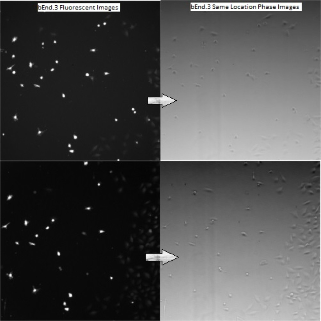
One thing that I’d done different from them in procedure, however, was that I would memorize positions on the Metamorph imaging software for specific locations within the wells, and therefore could take pictures at the exact same location multiple timepoints in a row. I began to notice that there seemed to be stunted growth in the bEnd.3 cells I looked at multiple times (each time exciting with ~498nm wavelength light), and even rounding up and possible cell death. Indeed, it appeared that cells were either rounding up (dying and detaching), or migrating to where the light was not being directed.
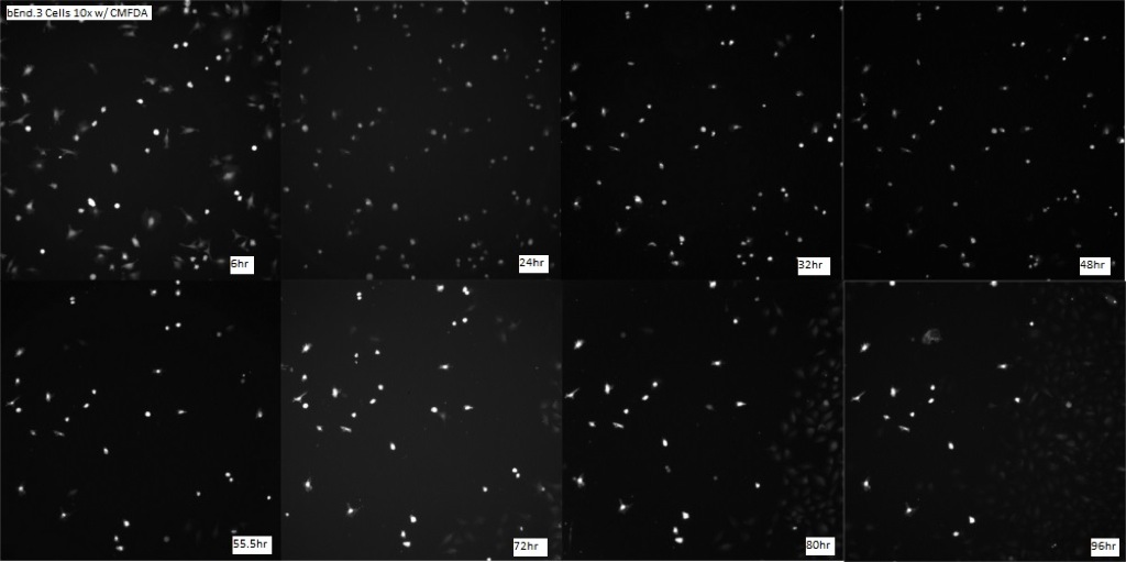
The growth curves showed the discrepancy in growth, with the areas that had been repeatedly imaged having cell populations which plummeted (some into single-digits), and even after more than four days still had only one-fourth the population of the locations without CMFDA. And the rebound in growth was not caused by the cells within the imaging window, but by other bEnd.3 cells which began invading from outside the viewing window and propagating along the side areas of the wells (see above images, especially the later timepoints).
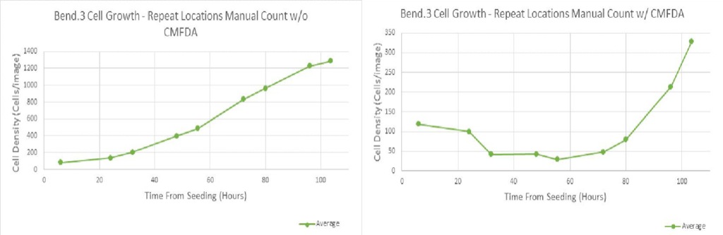
Part of the experiment also had locations which were excited once at the very beginning, then again at a specific time or times. This allowed for normalized numbers without consistent excitation of the cells. However, a similar effect was seen and the graphs showed a similar trend (where those areas with CMFDA had a heavy dip in cell population, while those without the stain propagated healthily and normally).
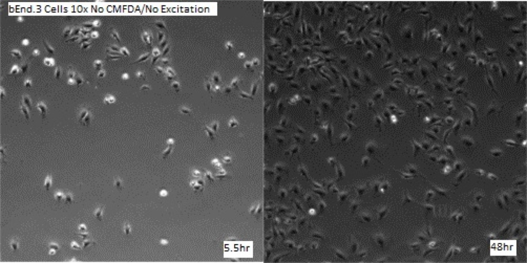
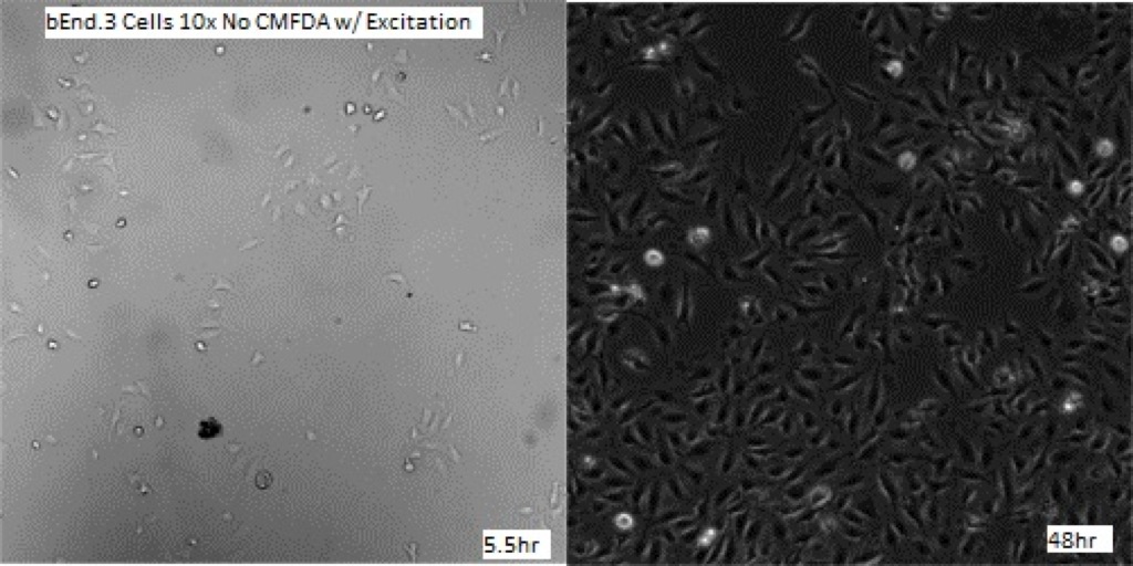
To evaluate what exactly was the cause of this negative influence on the cell’s viability, a secondary experiment was performed. Four wells were seeded with bEnd.3 cells, and two of the four had CMFDA stain added. One well with stain and one well without stain were solely imaged with phase and never exposed to the excitation light of the fluorescent bulb. The other two wells (one w/ CMFDA, one w/o CMFDA) were both excited with the ~498nm light for roughly 5 seconds each time they were imaged. The results appear to show that the CMFDA by itself was not causing the negative effect on cell growth when absent of the excitation light. Also, the excitation light alone did not seem to cause any issues. Only when there was CMFDA present and the fluorescent light was shined on it was there the detriment to growth seen.
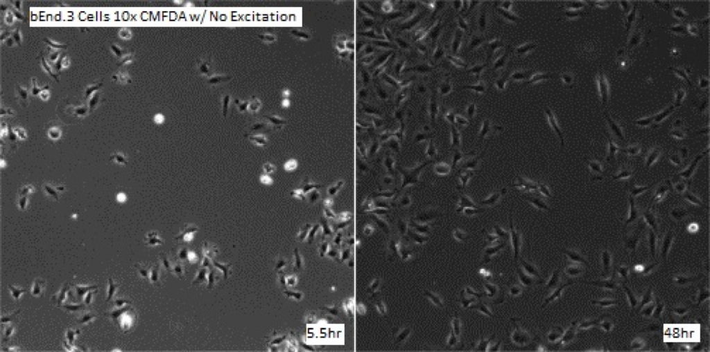
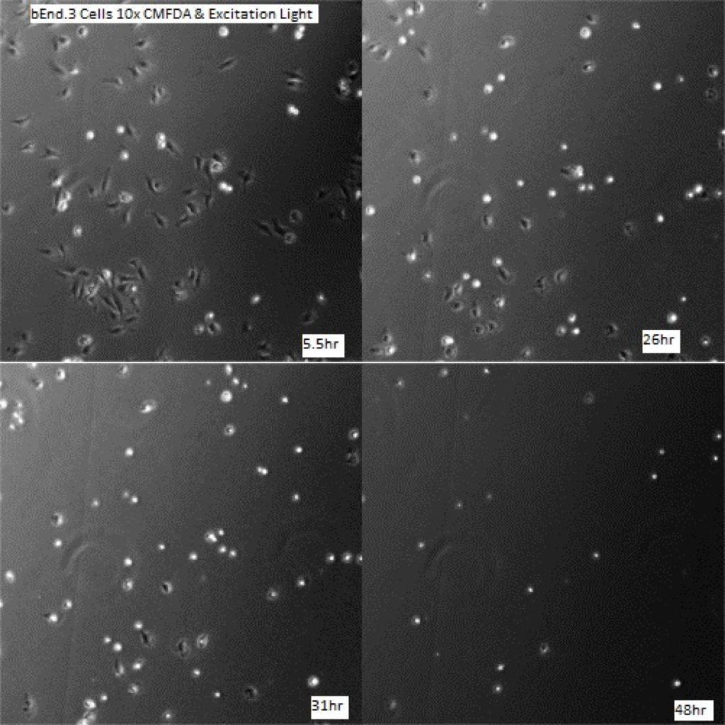
Continued research is looking into whether higher and lower concentrations of CMFDA (from 1uM ->25uM) have influence on this detrimental effect on bEnd.3 cells. In addition, HUVEC cells will be analyzed (in a manner of the final experiment here, with one control and three experimental groups) to determine the effect of CMFDA in that cell line. Due to their more fragile nature, it is likely that HUVEC’s will have an even more adverse response to the dye. The preliminary data from studying CMFDA in HUVECs has been collected, and is posted here.
Cells in general hate to be looked at with intense light, particularly towards the blue end of the spectrum.
It is good you tried exposing even your controls to the illumination, as that goes some way to controlling for the effect of the illumination.
You noticed that the dye is only toxic when illuminated – this is explicable since as fluorophores undergo photobleaching they produce free radicals which are toxic to the cells.
Two things to suggest if you can’t use a red-shifted dye:
* Try using less intense illumination and a longer exposure time (this will minimise the triplet state of the fluorophore and thus bleaching)
* Try incorporating an antioxidant/radical scavenger into your media – various compounds are used by for example the super-resolution community to minimise phototoxicity with intense illumination of fluorophores.