Co-Culture of ADSCs/HUVECs on 0.4um Translucent Membranes
Scope:
ADSCs were cultured on the bottom of the Corning 0.4um translucent membranes in DMEM+ 10% FBS+ 1% Pen-Strep. After 6 hours, the membranes were flipped over and placed into a 24 well plate. Of the four membranes with ADSCs cultured on them, 2 of them were seeded with HUVECs on the top side of the membrane. On day 3, two of the membranes were treated with CMFDA to ensure that cells were adhesive to the surface. This was done to one of the ADSC only membranes and one of the co-culture membranes. The CMFDA did not stain the cells well and cells were only faintly visible one day after staining. On day 8, the wells with CMFDA were stained with DAPI/phallodin (concentration of 1:100 for each), and the wells that did not have the CMFDA were stained with DAPI/CD31 antibody stain (CD31 concentration of 1:200). Images from after the cells were stained are placed below.
Results:
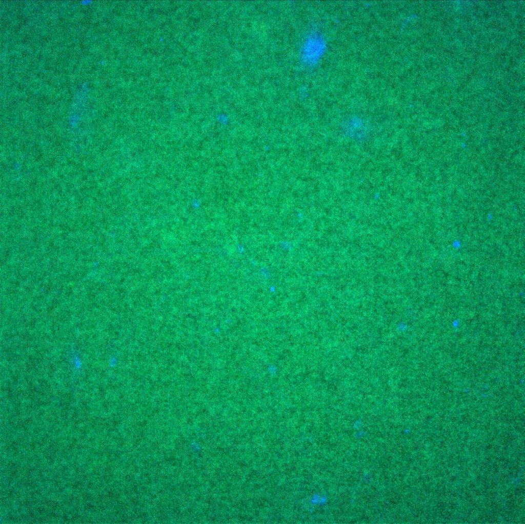
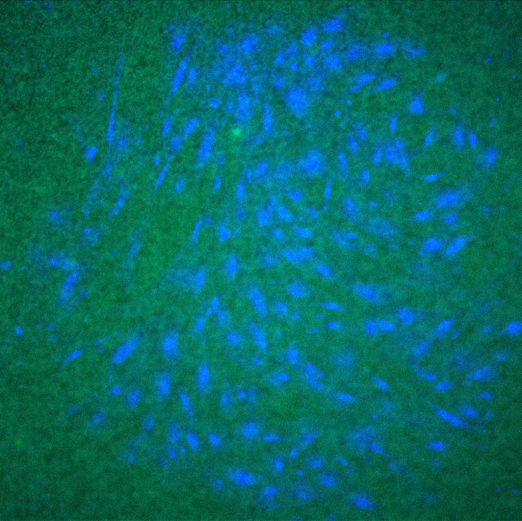
The cells that were stained with CMFDA and then the DAPI/phallodin both allowed for quality images. It appears that the morphology of the cells is quite different from the ADSC only sample to the co-culture sample. With the DAPI/CD31 stain, images did not turn out well. The nuclei stained blue with success but it appears that the CD31 stain was absorbed in the membrane and cell membranes were not apparently stained.
Images:
| Conditions | Images (20x) | Images (10x) |
| DAPI/Phallodin ADSC only | 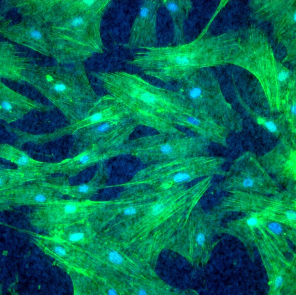 |
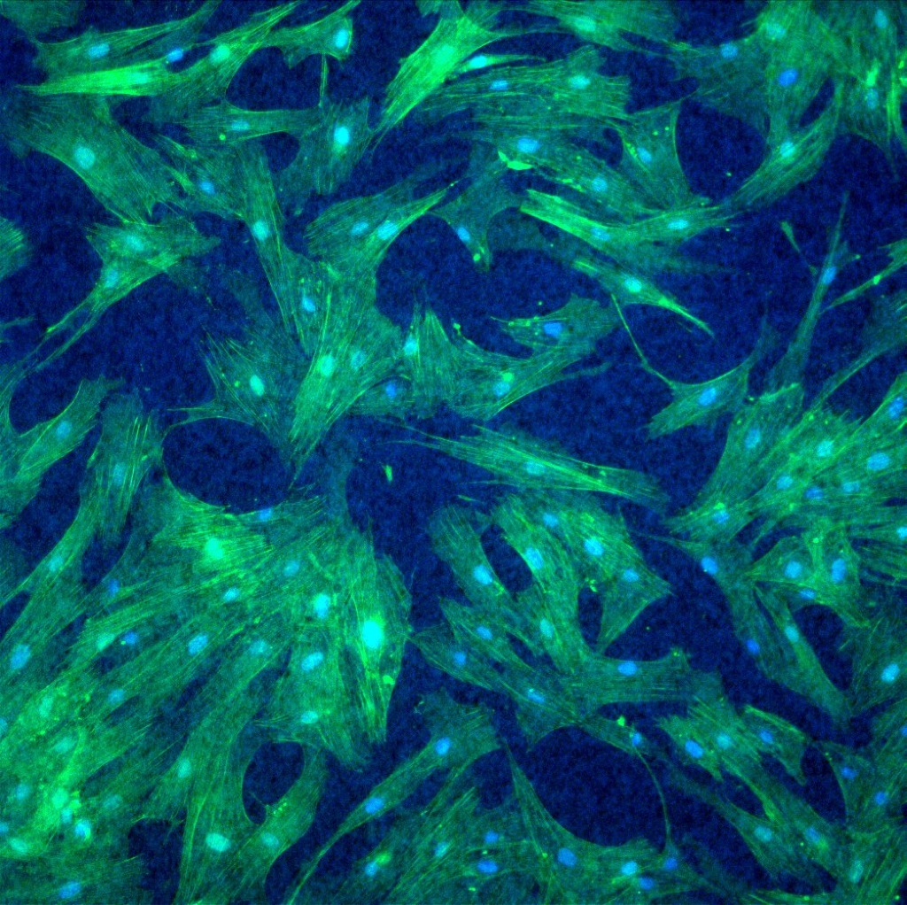 |
| DAPI/Phallodin Co-Culture | 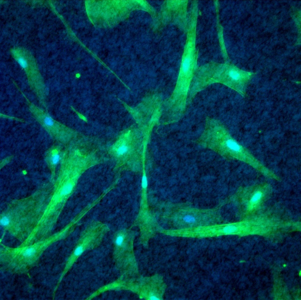 |
 |
| DAPI/CD31 ADSC Only | 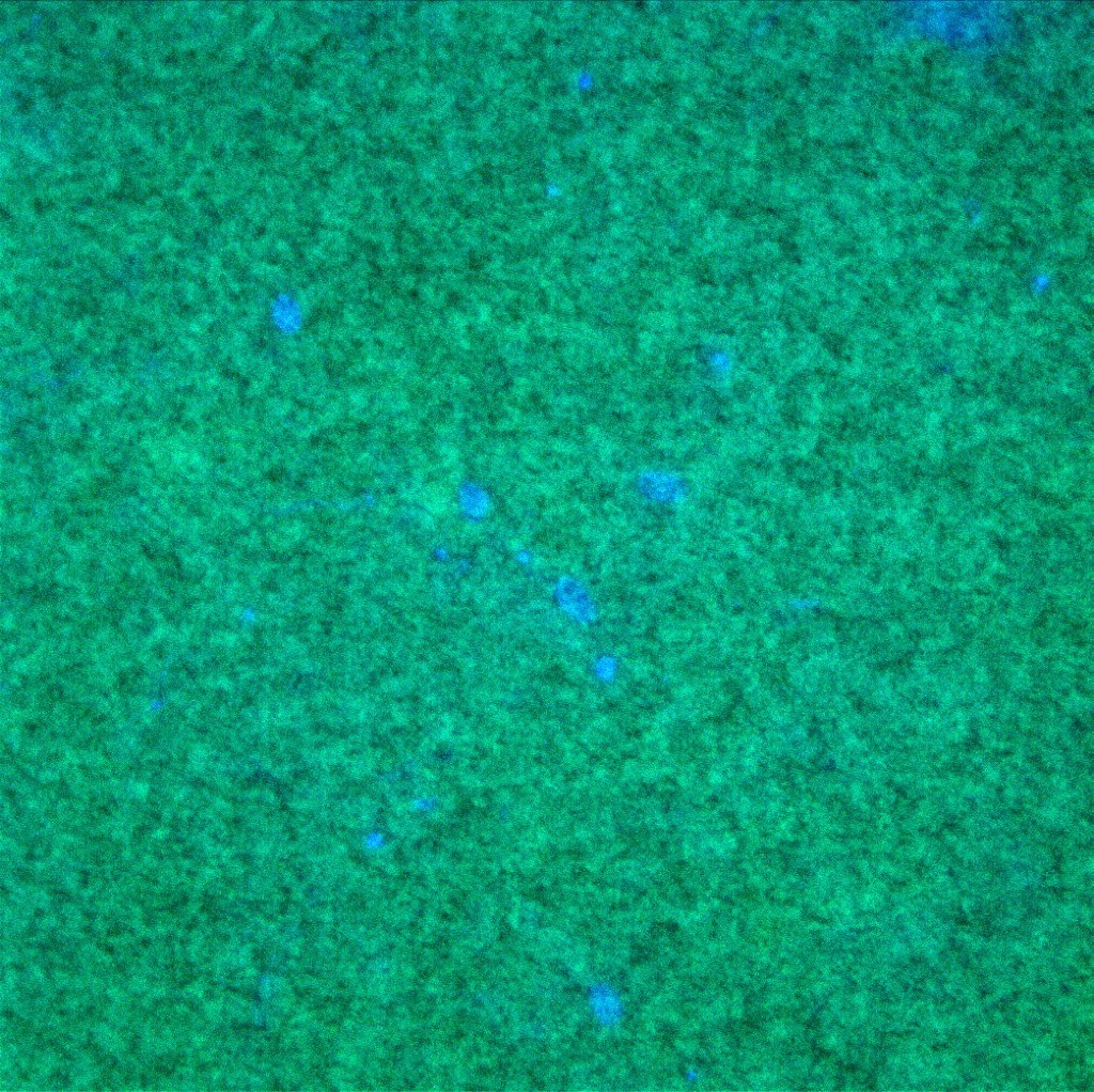 |
 |
| DAPI/CD31 Co-Culture | 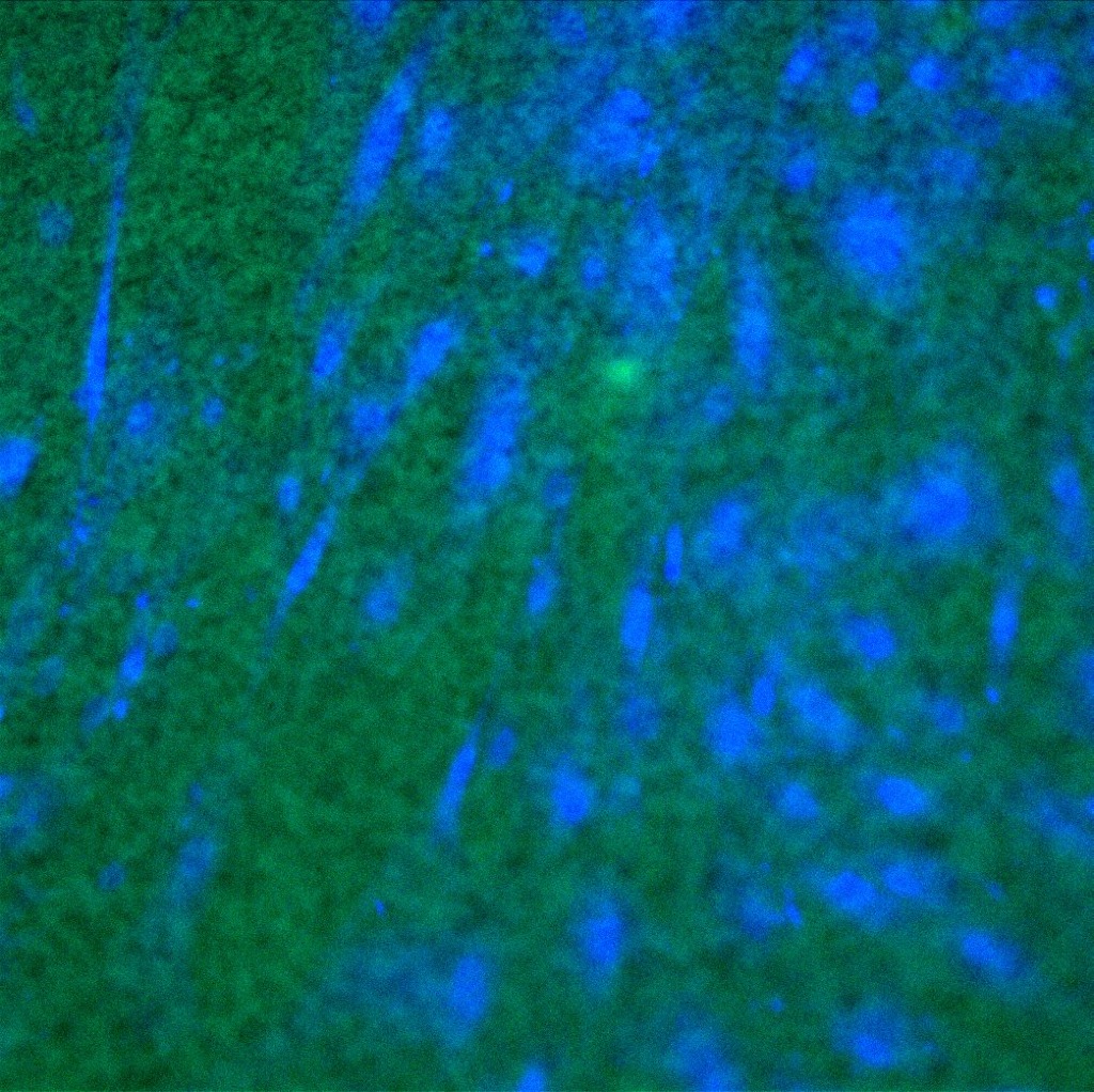 |
 |