Issues with cell growth on microporous membranes under flow
I am trying to grow endothelial cells under flow and measure electrical resistance of the culture during the flow-based growth. I am using a peristaltic pump to generate the flow. I am using microporous membrane with 3 micron pores with 3 micron spacing, and 20% porosity for this experiment. Microporous membranes can facilitate neutrophil transmigration, hence 3 micron pores. I assemble a co-culture device, with channels to feed both top and bottom chambers separately, and the design of the gaskets prevents any cross-communication of the liquid except through the membrane. I coat the flat side of the membrane with 1% geltrex to promote faster cell adhesion. The protocol has been optimized by Henry, and the only difference between his and my setup is the method of introducing the flow in the device. I am using a peristaltic pump where as he used a gravity feed setup. I have a small fluidic capacitor in my fluid circuit to reduce the pulsatile behavior of the flow introduced by the peristaltic pump.
After I seed the cells, I close the top and bottom chamber to prevent any kind of atmospheric convection happening in the device, and allow the cells to settle on the membrane uniformly and adhere and spread on it (I pre-equilibriate my media inside the incubator for an hour before I seed the cells, so that there is sufficient gas dissolved in it to serve the cells for few hours). After around 5-6 hours, I open both the top and bottom chambers to atmosphere, and keep the entire setup in a petri dish to maintain sterility. After around 24 hours, I connect the device to a pump, that derives the media from a PDMS reservoir, passes through the capacitor, flows through the device, and empties it back in to the reservoir. I take as many pictures as I can take to record the cell growth as time progresses.
As we can see from the following set of images, cells grow fine under static conditions for an entire day inside the closed microfluidic device. As soon as I start the flow, I see cells getting sheared and disappearing from the surface gradually. Comparing t=24 and t=25 hour images, one hour of flow reduced the number of cells by a significant amount. This phenomenon is very repetitive and I observed cell loss in almost every experiments. The shear stress is around 6 dynes/cm2, which is nowhere closer to shear the cells off.
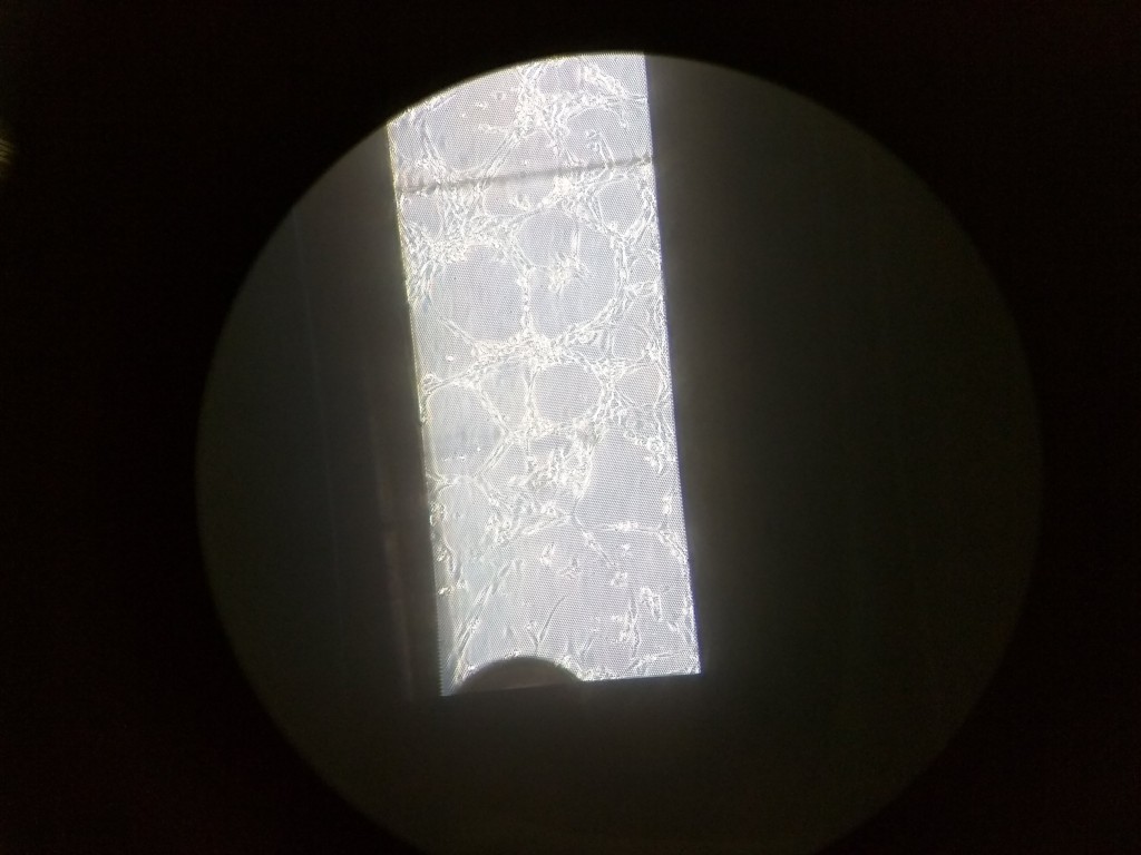
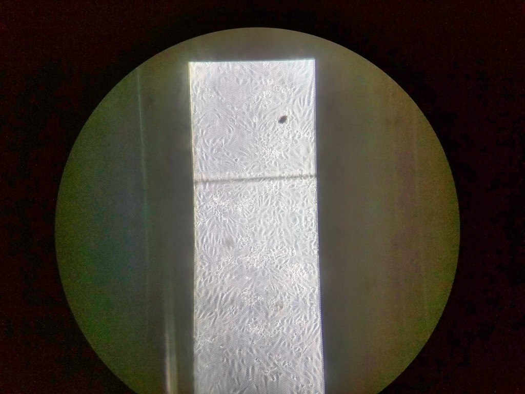
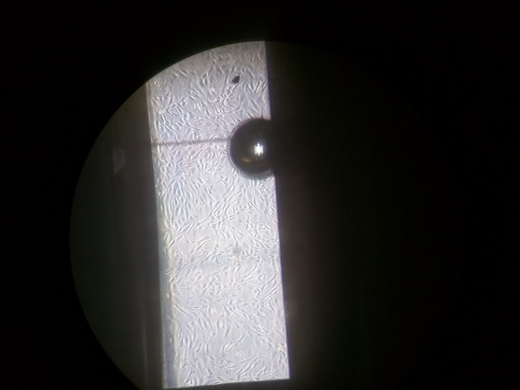
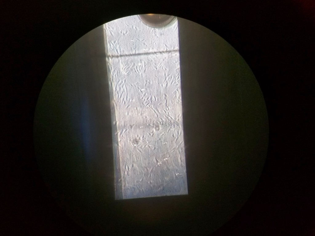
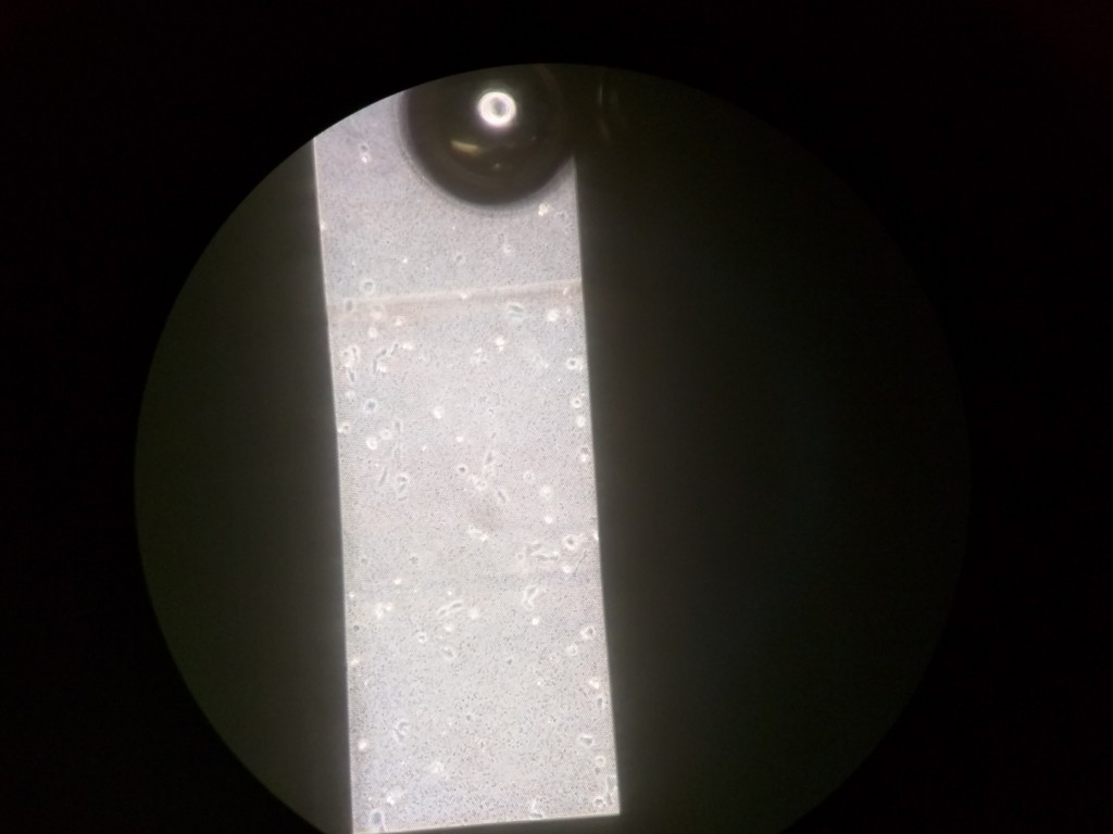
Our hypothesis is that, the membrane deflection is responsible for the cells to shear off. The flow induced deflection causes convection, and although the other side is not open, there is still media movement across the membrane pores. The pores effectively act as ‘jet stream’, and cells feel a pushing force from the bottom. Also from Henry’s paper on shear free device, we know that flow profile follows a parabolic behavior, with a maxima of flow coming from the bottom at the center of the membrane. Indeed in the past, I have noticed cells are completely gone from the center and only a handful of them are left at the corner. This membrane deflection is not necessarily due to peristaltic pump, and even the gravity feed can and will cause membrane deflection. The phenomenon is less prominent for nanoporous membrane, since the flow convection through the nanopores is lesser than micro pores.
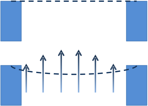
Furthermore, since there are 3 micron gaps after every 3 micron separation, cells have a significantly less area to adhere on to. Nanoporous membranes, in spite of similar porosity, have more substrate area to provide support, and cells can have better traction from below.
In order to rescue this experiment, I used a nanoporous membrane with 40 nm pores. Nanoporous membranes should exhibit less amount of this “jet-stream” phenomenon, and should be able to retain the cells for longer durations.
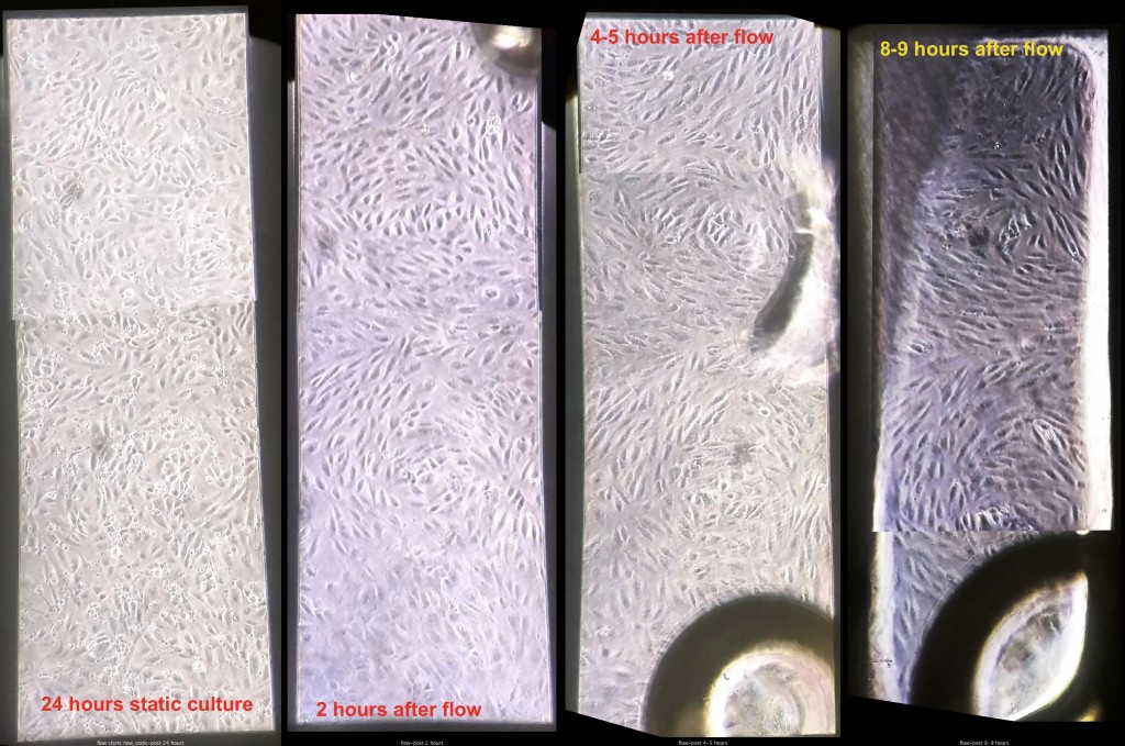
As we can see from the images, the flow induced alignment has circular growth patterns at both the ends of the channels within the visible area. It could be due to flow-induced membrane deflection or some other flow abnormalities inside the channel. The reason for this behavior is unknown, and has been noticed previously by Henry during his experiments.