Neural Crest Stem Cell Differentiation into Brain Pericyte-like Cells
Introduction
Breakdown of the blood-brain barrier (BBB) is one of the earliest signs of sepsis and is linked with long-term cognitive impairment. As animal models poorly recapitulate human sepsis, alternative approaches are direly needed to understand sepsis-associated brain injury and develop strategies that protect against cognitive decline. To fill this gap, we are developing a microfluidic model of the human blood-brain barrier (µSiM-hBBB) to elucidate mechanisms of BBB breakdown during sepsis. The first version of the µSiM-hBBB will be developed using human induced pluripotent stem cells (hiPSC) to derive two cellular components of the neurovascular unit: brain microvascular endothelial cells (BMECs) and pericytes. While BMECs are the main component of the BBB, pericytes are key support cell of the neurovascular unit (NVU), along with astrocytes and microglia. Pericytes not only help stabilize the BBB, but are key responders during inflammation and help guide immune cell transmigration.
Several protocols have been developed to differentiate pericytes from hiPSCs, however, only recently, a protocol was established to develop brain pericytes following the cells’ developmental pattern (Stebbins et al., Neuroscience 2019). This method drives hiPSCs towards neural crest stem cells (NCSC, p75-NGFR+HNK-1+) and then guides them into NG2+PDGFRβ+ “brain pericyte-like cells”. This post summarizes my first attempt at replicating this protocol in our lab, focusing on differentiation from NCSC into brain pericyte-like cells.
Methods
Pericyte Differentiation
D16
- The day following MACS, switch medium to Essential Medium 6 (E6) + 10% FBS (Peak Serum).
D17 on…
- Feed cells with E6 + 10% FBS daily. Pass when cells reach 90-100% confluency (Note: Cells become very difficult to singularize once they pass 100% confluency). Pass into uncoated dishes for final analysis (6 well plate for RNA extraction; 96 well plate for ICC; Transwells (Corning, 12-well, PET, 0.4 µm) for coculture permeability; µSiM devices enabled by NPN membranes (NPSN-100L, Wafer #1364) for coculture µSiM analysis).
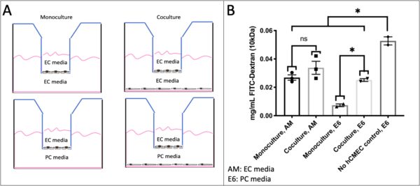
- For Transwell co-cultures, add pericytes to bottom chamber of Transwell at D19 and seed hCMEC/D3 at 20,000 cells/well in collagen-coated (Sigma) top chamber at D23. Half of the Transwells did not receive pericytes for monoculture controls. After adding hCMEC/D3, maintain in either all assay media (hCMEC/D3 medium) or assay media in the top chamber and E6 + 10% FBS in the bottom chamber.
- For device co-cultures (µSiM V1), add pericytes to topside of bottom channel at D19 (see ALine Modular Device Cell Culturing: Bottom Channel Culturing) and seed hCMEC/D3 at 40,000 cells/cm2 in top well at D23 (ALine Modular Device Cell Culturing: hCMEC/D3, Note I forgot to do 2 hour feed). After adding hCMEC/D3, maintain in all assay medium.
- For most devices, pericytes were seeded in uncoated devices. However, I coated three devices with 5 µg/cm2 of fibronectin (Corning) in case the pericytes did not stick to uncoated membranes.
- By D25, cells are ready for analysis. However, for coculture experiments, cells were maintained longer to allow hCMEC/D3 to grow confluent and mature junctions.
Analysis
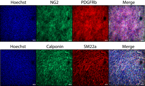
- D25: Fix cells in 96 well plate. Stain for pericyte markers PDGFRß, NG2, SM22, and Calponin following the methods in Stebbins et al., Neuroscience, 2019 (https://www.ncbi.nlm.nih.gov/pmc/articles/PMC6415958/).
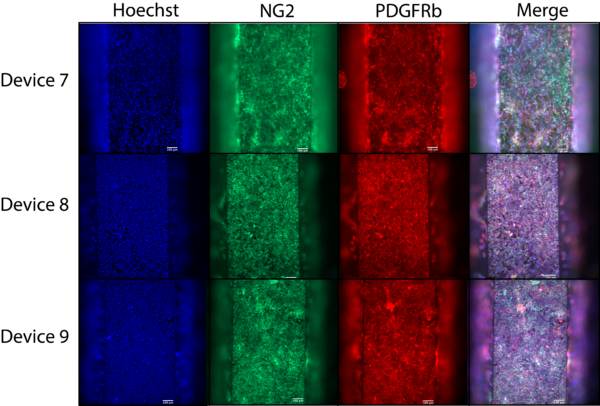
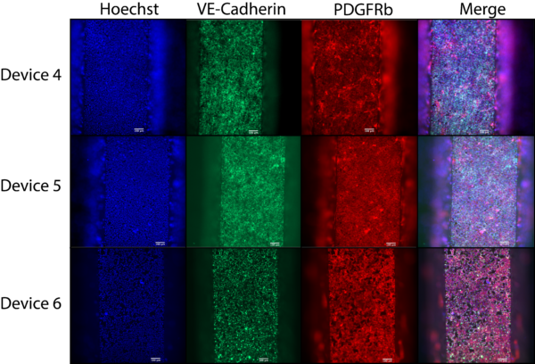
- D28: Fix cells in devices. Stain for pericyte markers PDGFRß and NG2, and endothelial junctional marker, VE-cadherin (https://www.ncbi.nlm.nih.gov/pmc/articles/PMC6415958/,ALine Modular Device: Immunocytochemistry).
- D28: Extract RNA from 6 well plates for qPCR and RNAseq (Qiagen, RNeasy Plus Minikit). A subset of cells were reverse transcribed (Superscript III, Thermo) and qPCR was performed (SYBR Green, Thermo).
- D29: Small molecule (10 kDa FITC-Dextran) permeability in Transwell. Add 1 mg/ml 10 kDa FITC dextran to the top chamber of Transwells and incubate 4 hrs. Collect media from bottom chambers and read fluorescence intensity. Use dye ladder to determine concentration of dye in bottom channel.
Results
Conclusions
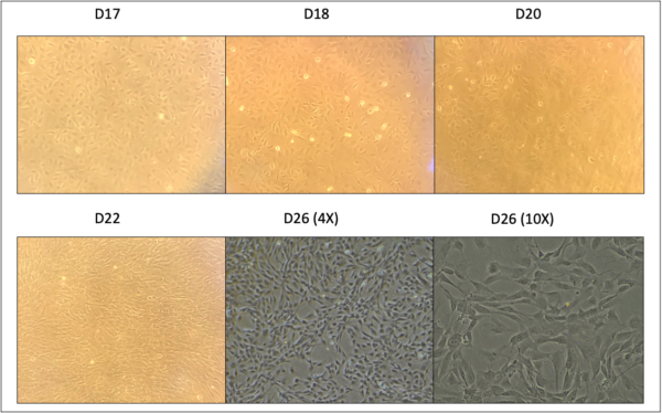
Based on preliminary analysis using ICC, we have successfully differentiated hiPSCs into brain pericyte-like cells. During the first 15 days of differentiation, cells were guided to NCSCs, with morphology matching Gastfriend et al (near submission?). Importantly, by day 12, we saw clusters of cells that were p75-NGFR–, which were subsequently sorted out via MACS. Flow cytometry analysis technique, while changing the percent of p75-NGFR+/HNK-1+, does not lead a difference in conclusion. We are still >90% double positive, which is the standard we set for “good enough” to move onto pericyte differentiation.
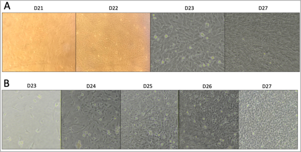
Following MACS, the cells were guided to pericytes, again with morphology matching Gastfriend et al (near submission?). It is important to note cells need to be passed prior to or right when they reach confluency, or they become extremely difficult to singularize and replate. This is especially important when subculturing into devices. In addition, although Transwells and µSiM devices were both seeded at 13,000 cells/cm2, the Transwells grew faster than cells in devices. Subculturing days will need to be optimized based on EC culturing. If pericytes are grown too long, the cells will peel off in sheets, so the ECs need to be ready (confluent with tight junctions) prior to that point. For hCMEC/D3, longer culturing is required than iPSC-derived BMECs.
At D25, pericytes subcultured in 96 well plates were analyzed for several pericyte markers, and the staining matched D25 staining by Gastfriend and colleagues very closely. This is our best indicator at this time for successful differentiation. RNA has been extracted from other pericytes, which will be used for RNAseq and qPCR of key genes for further analysis. qPCR is done but needs to be analyzed.
For µSiM culturing, it is unclear whether a coculture was successful, although ICC points towards success. After adding hCMEC/D3, I forgot to do a 2 hr feed to wash off dead cells. The next day, I assumed I was seeing pericyte growth and hCMEC/D3 had not taken, but it was impossible to distinguish between the two cell layers on our ultrathin membranes. Over the next few days, long processes appeared, while other cells formed a more cobble-stone appearance, as is characteristic of BBB cells. The cells in devices stained positively for both pericyte markers, NG2 and PDGFRß, and endothelial adherens junction marker, VE-cadherin. I did not have enough devices for proper controls. So, while this indicates both cells successfully grew together (iPSC-derived pericyte cells should not express VE-cadherin, as reported in Stebbins, et al., 2019), I would like to confirm further be separate analysis of the top and bottom of the membrane. In addition, while it is hard to tell, I am not sure the pericyte staining patterns match culture dishes, and would like to further analyze.
For the Transwell permeability assay, it appears pericytes actually reduce barrier function of hCMEC/D3 and cause an increase in small molecule permeability. While unexpected, this has been reported several times in the literature, while other reports indicate improved barrier function. Dr. Shusta’s group found reduced permeability when coculturing iPSC-derived brain pericyte-like cells with iPSC-derived BMECs. However, the coculturing time for those experiments was 24-28 hrs, whereas hCMEC/D3 take longer to reach confluency and form tight junctions, and, as such, were cocultured for 1 week. The trend was consistent between both medias, indicating it is likely real within our conditions and cell types. The other interesting takeaway is hCMEC/D3 actually perform much better when cultured with E6 + 10% FBS (PC medium) in its bottom chamber, regardless of whether they are in a monoculture or coculture. It is unclear why this is, as most reports show serum concentrations above 5% reduce barrier function. It may be a directional response of the cells, as that media is only contacting the cells from below. Maybe the reduction in barrier function in coculture has to do with pericytes using up the media components that are helping the hCMEC/D3 improve function, however this is just conjecture and not important for our purposes. For our purposes, we have shown that we can successfully culture iPSC-derived brain pericyte-like cells with endothelial cells and modify function, and while the functional change is not in the direction expected, I do not plan on exploring this further, as we are more interested in how our iPSC-derived endothelial cells will respond to pericytes. If we see the same phenomenon, then we may have to add astrocyte conditioned media in our system to get proper barrier function.