No Time To Waste: Developing a Faster Method for Viral Genome Extraction from Waste Water Samples
The premise of this project is built around the ideas that our membranes can:
- Be used as concentrators for very dilute targets
- Catch and hold particles for further processing on the membrane
Here, we wish to test these ideas for dilute viral genome extraction in large volumes of waste water. We looked to improve upon the protocol Direct wastewater RNA extraction via the “Milk of Silica (MoS)” method – A companion method to “Sewage, Salt, Silica and SARS-CoV-2 (4S)” by using our membranes and a vacuum filtration set up in lieu of multiple centrifugation steps and extremely high silica bead concentrations. Discussed below, we were able to successfully modify the protocol to make several improvements, but were unable to get conclusive results in our DNA extraction.
Protocol Modifications
Washing Buffer Contents
To make the washing buffers, we made our own 5 M NaCl solution (added 14.61 g of NaCl to 50 mL of DI water) and our own Tris-EDTA solution (added 90 uL of 1 M Tris-HCl pH 7.4 to 10 uL of 0.5 M EDTA). We also scaled the total volume of each solution down to 100 mL.
Use of Vaccinia Virus
As opposed to using a RNA virus as is done in the original protocol, we decided to try extracting DNA from vaccinia virus. This is because we had both whole vaccinia virus and purified DNA with a corresponding qPCR assay and reagents on hand from previous work. We extracted DNA from 1 copy/mL, 10 copies/mL and 100 copies/mL of whole vaccinia virus and used pure vaccinia DNA at the same concentrations as a positive control.
Membrane Type
We used 8 um slit pore membranes for these experiments.
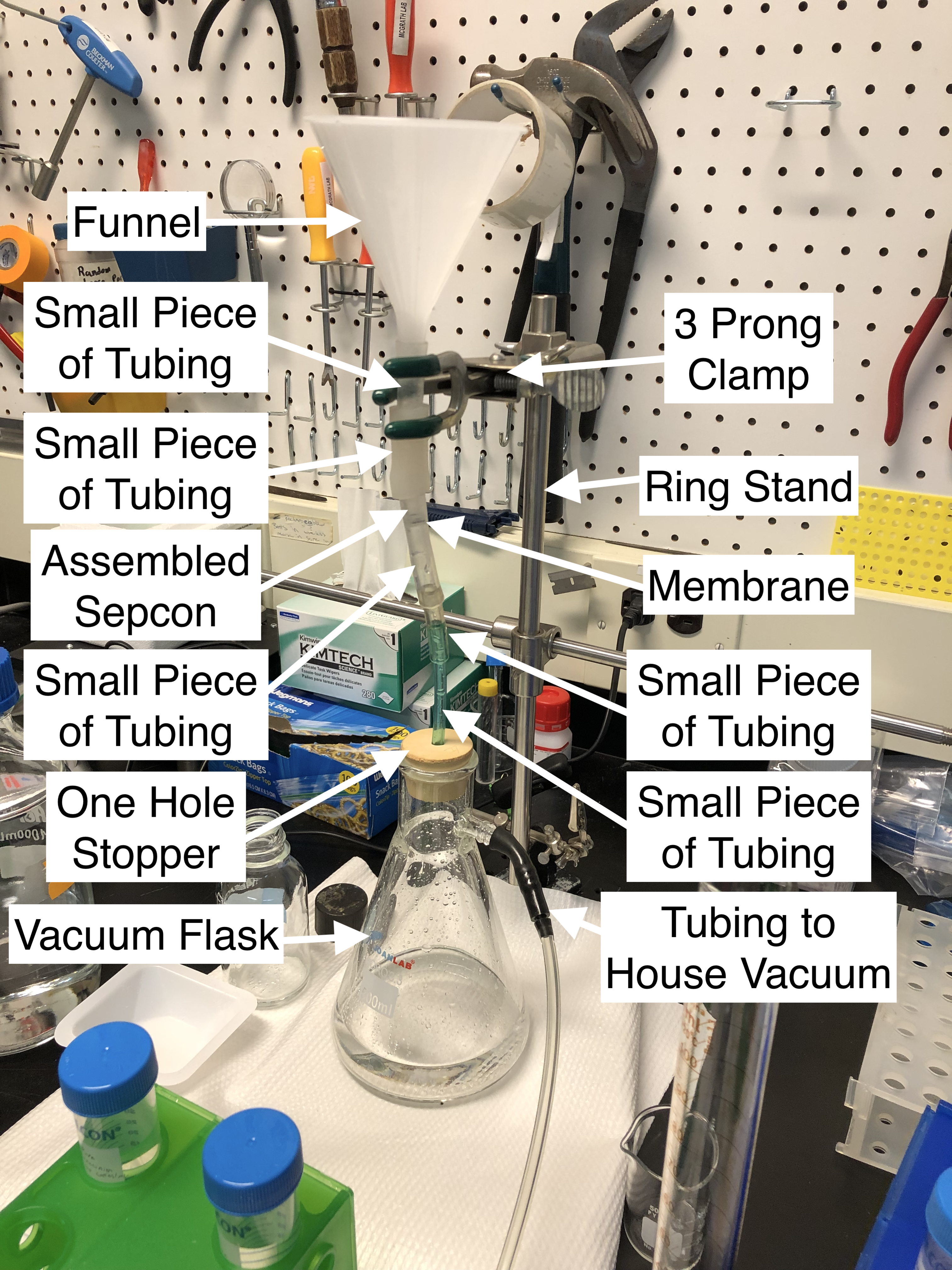
Vacuum Setup
Shown below, our vacuum setup comprised of several components including:
- a funnel
- several pieces of tubing
- a ring stand
- a 3 prong clamp
- an assembled sepcon containing an 8 um slit pore membrane
- a one hole rubber stopper
- a vacuum flask
- our house vacuum
Once assembled, we added solution to the funnel and filtered it through the membrane and into the vacuum flask. Once capture was complete we disassembled the setup, retrieved the sepcon and membrane, and washed and dried each component before re-use.
Silica Bead Concentration
Instead of using billions of 1-5 um diameter beads for our milk of silica solution, we used 1 million 10 um diameter beads. This ended up clogging the membrane rapidly to the point of stopping filtration, so we reduced the total number of beads that we used first to 100,000 beads, then to 50,000 beads.
Modified Protocol
For our DNA extractions, we modified the original protocol shown above to the following:
For virus lysis and DNA stabilization
– Dissolved 9.5 g of NaCl into 40 mL of diluted vaccinia DNA – Added 400 uL of Tris-EDTA solution
– Inverted 10 times and let sit for 10 minutes
To attach DNA to the silica beads
– Diluted the sample in half (80 mL total volume) using 70% ethanol and mixed thoroughly
– Added milk of silica solution (using a volume that corresponded to the desired total number of beads described above)
– Inverted 10 times and let sit for 10 minutes
To capture the DNA bound beads
– Added to vacuum setup and applied vacuum until all fluid was through, then equilibrated the setup to atmospheric pressure
To clean the beads of unwanted viral debris
– Added 1 mL of 4S-WB1 to the sepcon and let sit on the membrane for 5 minutes
– Re-engaged the vacuum to suck all fluid through, then equilibrated the setup to atmospheric pressure
– Added 1 mL of 4S-WB2 to the sepcon and let sit on the membrane for 5 minutes
– Re-engaged the vacuum to suck all fluid through, then equilibrated the setup to atmospheric pressure
– Repeated the previous two steps to wash the membrane once more with the 4S-WB2 solution
To release the DNA from the surface of the beads
– Extracted the sepcon from the vacuum setup and plugged the holes of the bucket with parafilm
– Added the desired volume of nuclease free water (50 uL) to resuspend extracted DNA and incubated for 10 minutes
Setup Performance
Overall, the setup worked as we had expected. Vacuum would suck fluid through the membrane easily until beads began capturing on its surface. We learned that we initially had too many beads in solution, over saturating the membrane, forming a visible cake and clogging it completely. In response we decreased our bead concentration twice until we got to a point where we thought complete membrane occlusion would not occur. Throughout testing the membranes would remain intact unless we left the vacuum at full power when the final portion of fluid was sucked through. Therefore the setup seemed most liable to destruction via its air/water interface. Nonetheless, we were able to complete our protocol in ~1.5 hours compared to the ~6 hours that the original protocol requires.
qPCR Results
After extraction, we had a total of 3 unknown samples to quantify. We tested these alongside positive controls of purified vaccinia DNA and negative controls containing just water, master mix, and primers. All amplification was compared to a standard curve which had a range of 10^0 copies of DNA to 10^7 copies of DNA. Unfortunately, almost every unknown sample and positive control amplified at the same level as or beneath our water negative controls. This meant that the data was inconclusive due to the unknowns and positive controls being at or below the sensitivity of the qPCR assay. Even with perfect recovery for the highest concentration of 100 copies/mL in a 40 mL sample, this means there is a total of 4000 DNA copies in our final extraction volume of 50 uL. Injecting only 5 uL of this solution means that our qPCR assay is trying to detect 400 total DNA copies which is near the lower limit of detection of the assay.
Addendum
While reviewing these results and preparing for future experiments, it seems that we mistakenly used polystyrene particles and not silica particles. This would explain why our results were so poor.
Future Work
To truly determine whether or not we are successfully extracting DNA from solution while using this same protocol, we need to increase the concentration of vaccinia DNA to get within a range that can be detected with our qPCR assay. We now know the bottom limit of the assay is ~10^2 DNA copies, so extracting a range of concentrations above this should provide results that we can use to gauge the effectiveness of our extraction protocol.
Does the nuclease-free water do the extraction? What was the ‘desired volume’ you used in the this step. Isn’t that the volume that determines the concentration in your assay?