Immunogold Identification of Exosomes
This is pretty much an update to a previous post on trying to identify exosomes using an immunogold staining procedure, FIB lift-out and the TEM. After multiple attempts to cut out a section of the membrane for attachment to a TEM grid (we were able to cut it out and weld it to a grid the first time, but in the transition between the SEM and the TEM, the sample was lost), we were finally able to get a segment of the membrane that survived from the SEM to the TEM. We can see in Fig. 1 that the membrane is welded to a side of the copper mesh grid and there are four platinum support structures to prevent the membrane from folding in on itself.
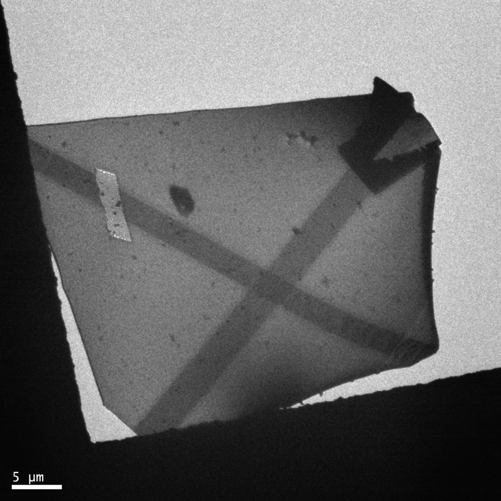
Figure 1: TEM image of the membrane piece welded to copper TEM mesh.
Upon closer inspection, there was a lot of non-specific bonding of gold nanoparticles (the gold conjugated secondary) to the membrane itself. However, there were particles that appear to be exosomes also with gold particles localized to them, as can be seen in Fig. 2 and Fig. 3
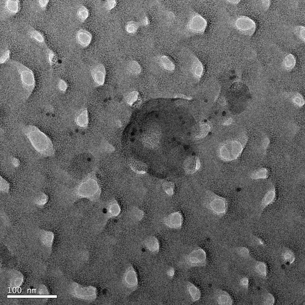
Figure 2: Gold particles binding non-specifically and around a lipid vesicle. The vesicle is stained with uranyl acetate for contrast. The white spots are pores.
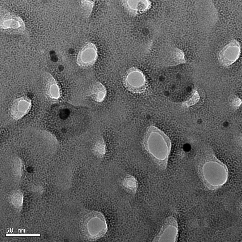
Figure 3: Gold particles localized around two exosomes with non-specific binding in the region.
As we can see, there is a decent amount of non-specific binding present in these samples, which arises for a reason that I will postulate in just a minute. However, I do want to note that there appears to be higher concentrations of the gold particles in the region of the exosomes, which would suggest specific binding. This is also observed in my previous SEM image of the surface after antibody conjugation, shown in Figure 4.
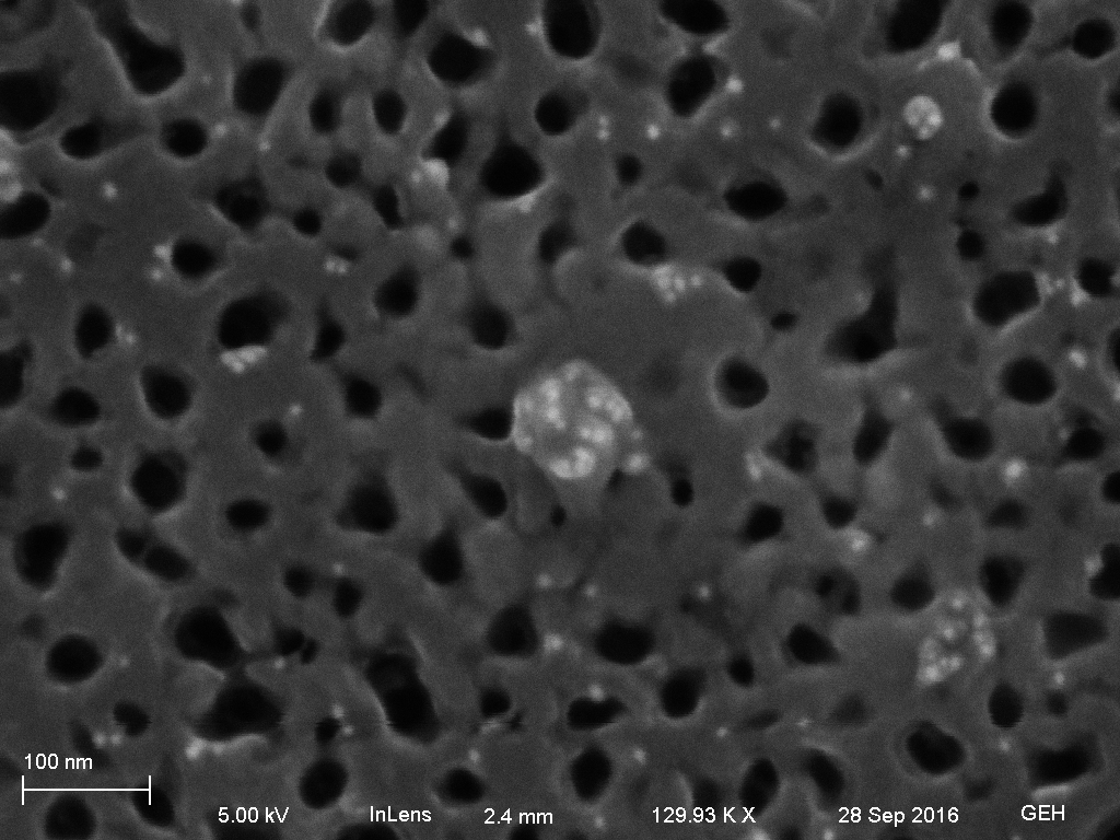
Figure 4: SEM micrograph of antibody treated surface showing a particle covered with gold nanoparticles, with non-specific binding of gold particles elsewhere on the surface.
This brings me back to a point that I mentioned earlier. In the TEM, our exosomes appear different than common images of exosomes in the literature. Additionally, TEM micrographs of exosomes in the literature show very little residual gold nanoparticles in the sample. My hypothesis for this is related to the preparation procedure that is significantly different for both samples. In the literature, exosomes are isolated and then suspended in a droplet, on to which a TEM grid is placed. The liquid exosome sample is then suspended in the spaces between the pores and fixed there. The residual gold nanoparticles can then be washed out of the space between the exosomes, so there is no background. However, I am preparing these samples directly on the membrane and therefore non-specific binding is allowed and will occur. Even with rigorous washing, there will still be gold bound to the surface that will show up in the TEM images as the non-specifically bound gold. Additionally, our exosomes will also appear slightly different due to the imaging interference from the membrane itself. This is a good first step in this process of identification, but the next step will be to perform a control with PBS instead of plasma. If we can show that there is non-specific binding to the surface of the membrane, but it is not localized as we see around the exosomes, then this should be more proof that we are indeed seeing exosomes.
I want to leave you with a final image that I believe shows the exosome/gold conjugation quite nicely. There is a gold particle sitting a short distance away from the exosome, in the same plane. This small separation should be the antibody that the gold is conjugated to and we tried to image the antibody itself, but that proved to be too difficult.

Figure 5: Exosome and gold particle in the same plane (not on top of each other) possibly confirming that there is an antibody connecting the two.