Adenovirus Captured and Imaged
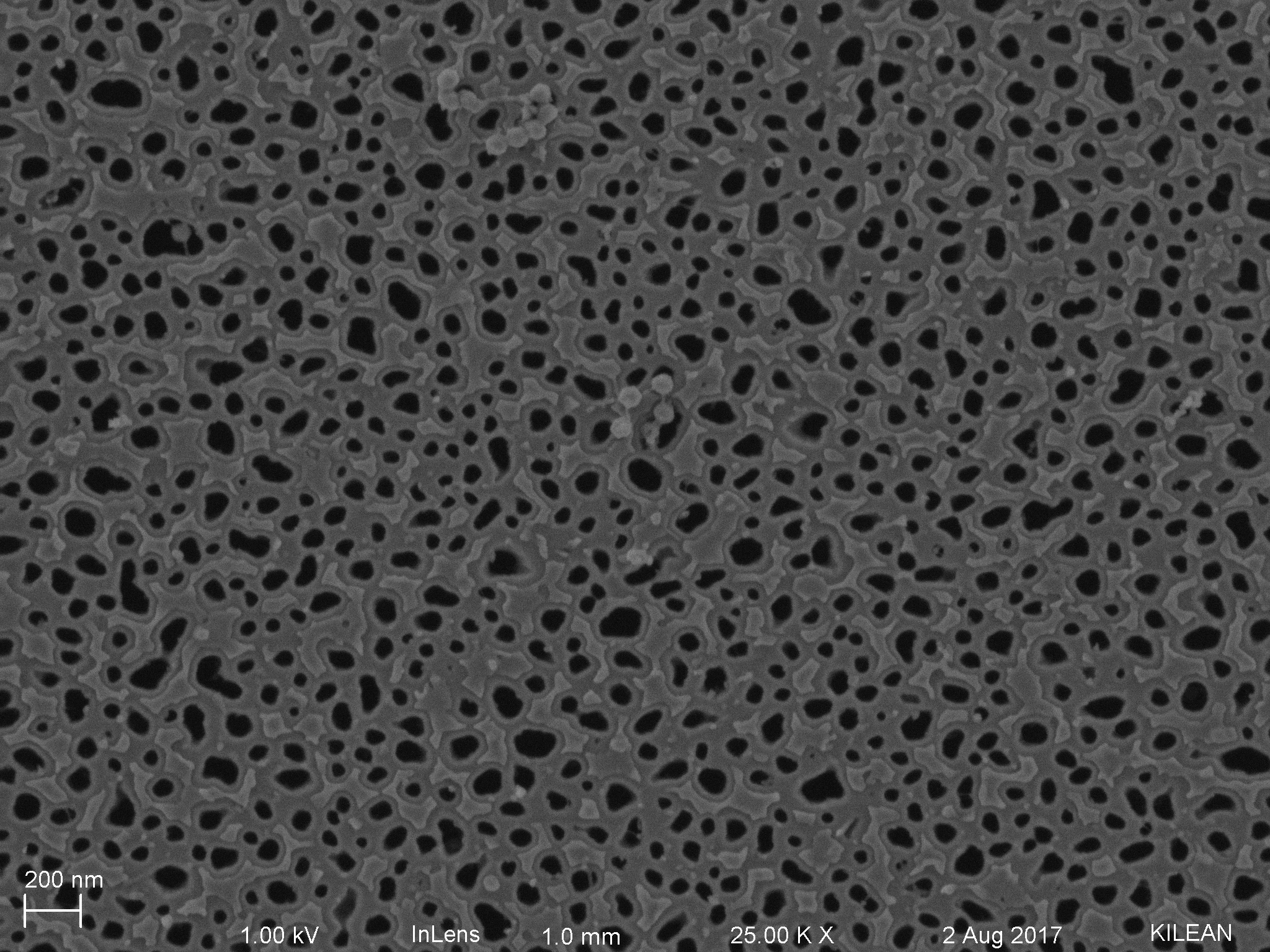
Last week we were able to capture and image individual virions on the nano membrane without a PETL using a simple drop-on-membrane protocol to establish some pull through. This was done to to verify that the individual virions will actually be caught in the pores and that our EM preparation protocol was efficient for drying the sample without harming it. The next step is too run the same experiment throughout the PETL dialysis system that captured gold particles and mimics Kilean’s tangential flow work to hopefully produce the same results.

Future Directions for Improvements:
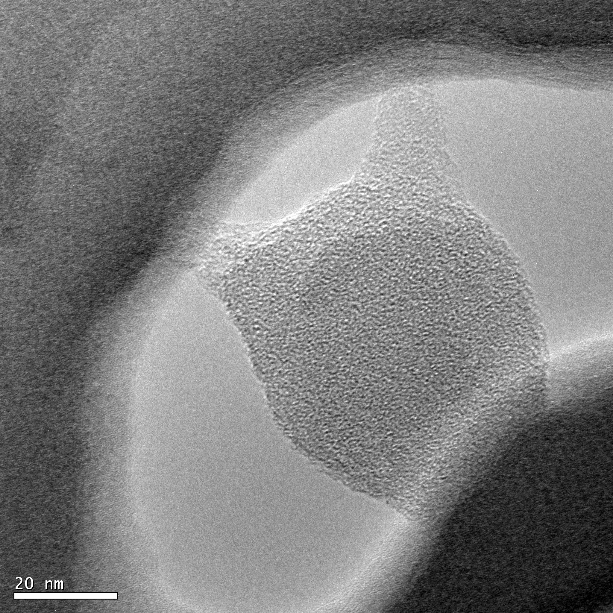
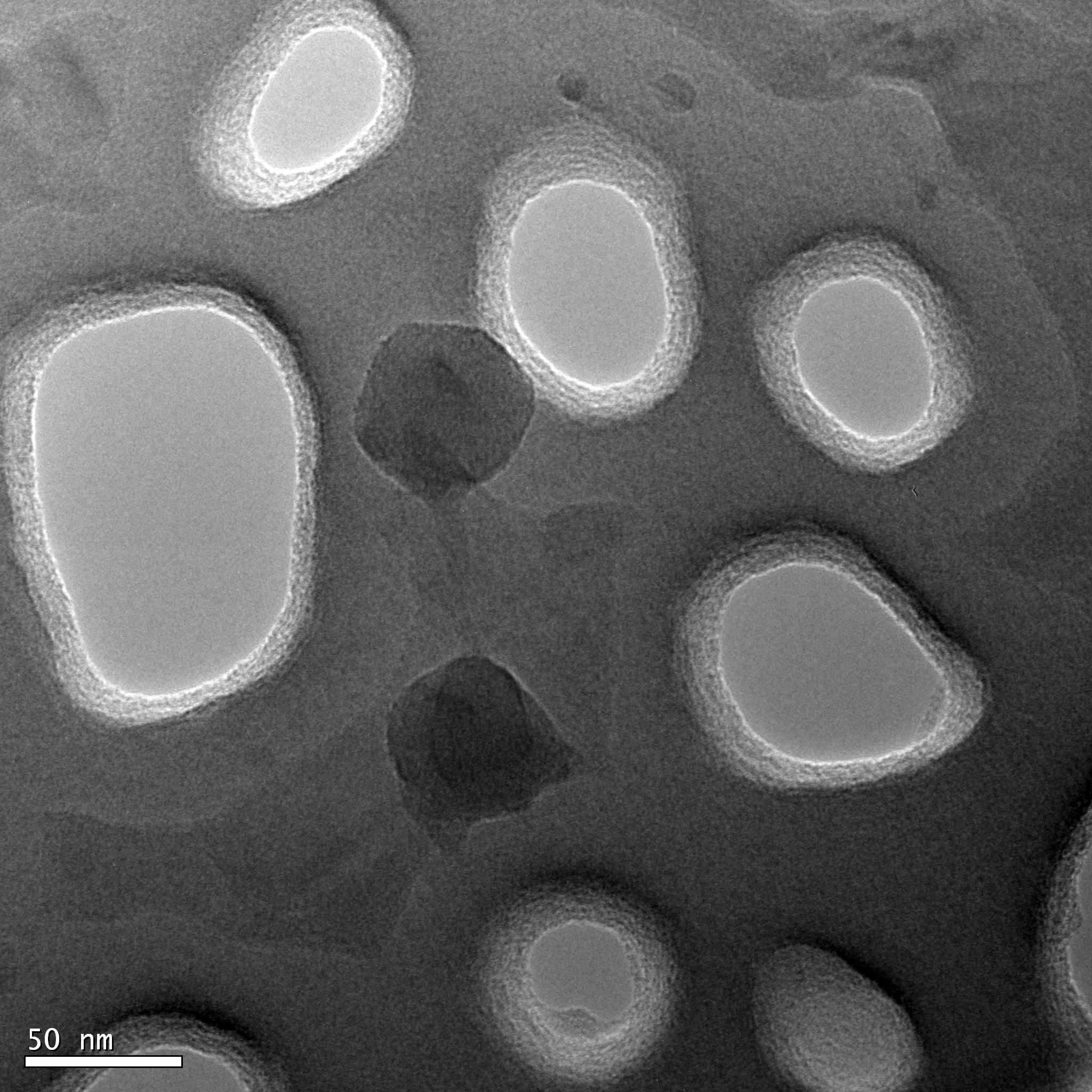
Currently only some Icosahedral structure can be observed throughout the sample which does not help when trying to verify that this is actually adenovirus caught in the pores.
- Using a fixative (glutaraldehyde) prior to dehydration might allow for structure to be preserved by cross linking the amine groups in certain proteins.
- Using a Cryo-EM preparation method and a computational 3D reconstruction method to embed the sample in vitreous ice and help determine the structure from TEM imaging.
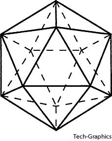
- Viewing the particles from different angles might also help reveal some structural components such as the pentose bases (corners) due to its icosahedral shape giving it a somewhat circular shape.
- potentially producing a tilt stack.
- Ultra-centrifugation might help separate individual virions from each other and from the buffer they are stored in (10mM Tris-HCl (pH 7.5), 1mM MgCl2, 10% glycerol) which may cause some imaging errors.
- Coating the sample with more gold platinum to produce more of a shadow around the particle might make the morphology more defined.
- Using a positive stain on the sample might also allow for better contrast when imaging for structure inside a pore.

Awesome but … Adenovirus doesn’t look like this in other electron micrographs? https://www.vetarena.org/gallery/image-1042.l.jpg
In the second picture of the virion on the TEM i believe it is stretched out due to some adhesion or interaction with the wall of the pore when going through the drying process. The first image is a little zoomed out but if you look in the center there are some virions that seem to be attached together that show the icosahedral structure we are looking for. Why they are attached together, I am not completely sure. I do not see any of the fibers that should be sticking out of the nucleocapsid, as seen in the image you attached. This could be due to the way it was prepared in the lab prior to us receiving it or it is probably due to the microscopy. I had a chat with a virologist here at fisher and her input was that these fibers are so thin and short (about 5nm extrusions and 1-2 proteins wide) that they might not be able to be seen in the resolution or visibility range that we used to image. There are a few more factors that could have caused this and it would be impossible to point out exactly the reason, but it would be too easy if everything worked the first time!