Beadazzled: Laying the Foundation of the μSiM-DX Fouling-based Sensor
Starting in December of 2019, we began capturing fluorescent beads on microslit silicon nanomembranes as an illustrative representation of particle capture within our microfluidic devices. From then until now we have successfully used this method within gasket made TFAC devices and uSiM ALine devices using a range of micropore sizes/shapes and bead sizes. These experiments have provided evidence for many anecdotal lessons over the years and have also been used to optimize a µSiM based sensor. These findings are elaborated on below.
Bead Capture in the TFAC System
*Sped up and compressed videos for each tested chip condition is available in the appendix at the end of this post
Using a traditional Tangential Flow for Analyte Capture (TFAC) setup (simplistically, a microfluidic device with a top and bottom channel separated by a silicon nanomembrane), Kilean and I used 1 µm slit pore membranes in clamped devices to capture 1.24 µm diameter streptavidin coated polystyrene beads. The goal of our experiments were to prove the presence of Kode biotin on the surface of our membranes and to show that this surface bound biotin could readily interact with streptavidin. To test this we used streptavidin coated beads in a flow regime with three distinct steps: 1-Initial pore fill (2-3 minutes long), 2-Bottom channel pressure release (1 minute), and 3-Reverse flow (2-3 minutes). The purpose of step 1 is to allow for bead capture, while the purpose of steps 2 and 3 is to release beads which are not captured via affinity. In this manner, beads that are only physically captured should be released and beads that are captured via affinity should stay on the membrane. Using an injection flow rate of 25 µL/min in the top channel and a withdrawal flow rate of 12.5 µL/min in the bottom channel with 1mL of a 1E7 beads/mL solution, we were able to successfully accomplish our goal. Repeatedly, biotin coated membranes were able to capture and hold onto streptavidin coated beads better than bare membranes. These observations were made with 1 µm and 8 µm slit pore membranes using 1.24 µm diameter streptavidin coated beads, shown in Figures 1 and 2 respectively.
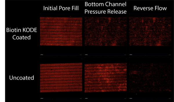
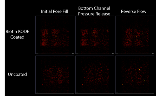
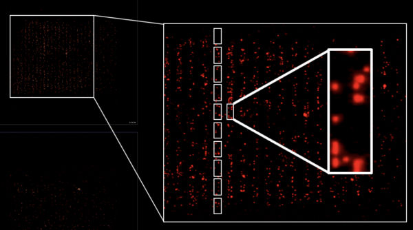
In both Figures it is shown that uncoated and biotin coated membranes initially capture beads similarly, but a noticeably larger amount of beads are able to remain on the coated membranes after steps are taken in our flow regime to take them off. The 1 µm slit pore membranes provide an example of capturing beads physically, but anchoring them to the membrane via an affinity-based interaction. The 8 µm slit pore membranes provide an example of purely affinity-based capture. Both scenarios provide evidence for biotin-streptavidin interaction on the surface of our membranes. Interestingly, a closer look at the 8 µm slit pores reveals evidence for the previously anecdotal idea that flow in the pores of our membranes “focuses” particles towards the sidewalls of the membranes. Shown in Figure 3, beads can be seen clearly lining the pores of the 8 µm slit pore membrane.
Within both sets of data, we noticed a nonzero baseline represented by the beads which were still able to hold on to the membrane in the absence of specific affinity-based capture. We attributed this to nonspecific binding with the membrane due to intermolecular forces, potentially caused by the streptavidin coating on the beads. In an attempt to reduce this baseline, we tried a new round of testing with bare 1 µm slit pore membranes that were coated with Kode PEG or BSA. Unfortunately, both treatments did not decrease the baseline that we saw and instead, they both increased the amount of beads that were captured nonspecifically. This may be due to the PEG or BSA presenting a polymer or protein “forest” on the surface of the membrane which provides the opportunity for streptavidin coated beads to get entangled and captured on the surface of the membrane.
To see if streptavidin coated beads would capture on the surface of a biotin coated silicon membrane without tangential flow, we coated nonporous membranes in biotin and used the same system and method as above. These experiments showed that tangential flow was necessary for bead capture, at least on a minutes timescale, after no beads were captured on the biotin coated nonporous membrane.
Bead Capture in µSiM ALine Devices
*Bead capture videos shown in the appendix
In µSiM ALine devices, beads have been used to optimize a fouling-based sensor. The microfluidic design of this fouling based sensor consists of an injection port and two outlets: the well and the indicator port. Connecting these are channels with their own independent resistances. The silicon membrane occupies the path leading to the well and has a variable resistance which depends on the degree of fouling. The device, which we call the μSiM-DX (microfluidic device featuring a silicon membrane for diagnostics), is based on the μSiM platform originally designed for tissue chip applications. In its test ready state, the bottom channel is pre-wet, and the well is partially filled to establish fluid continuity throughout the device. When sample without beads is injected, the membrane pores remain open and the flow path leading to the well is the path of lowest resistance. When the pores are clogged with beads however, the membranes act as a ‘resistance switch’ to redirect flow to the indicator port. This produces a visual positive in the form of a bubble that protrudes from the indicator port.
Initially beads that were larger than the pores of the membrane were used in order to determine which versions of the μSiM-DX operated properly. Testing included the use of 0.5 μm/1 μm slit pore membranes and 0.5 μm/1.24 μm diameter streptavidin coated beads respectively. Moving forward towards testing the affinity-based linker system of the sensor (between the biotin coated surface and the streptavidin coated beads), we also tested with streptavidin coated beads that were smaller than the pores (200 nm diameter in size). Shown in Table 1 and Figure 4, we were able to use the data from these tests to determine which sensor design would produce the most reliable and accurate results.
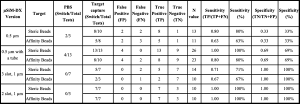
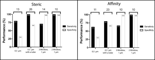
This data guided us through sensor designs. The first sensor we tested used 0.5 μm slit pore membranes. This design produced too many false positives and negatives. Most notably false positives were produced when testing with just PBS. This led us to believe that the resistance of the membrane was too high in comparison to the bottom channel of the μSiM-DX. In an attempt to address this, we added PEEK tubing (750 μm inner diameter) to the indicator port of the μSiM-DX to increase the resistance of the bottom channel. Ideally this would make the resistance of the bottom channel closer to the resistance of the membrane, allowing the device to perform better. This adjustment produced better performance overall, especially with steric capture, but there was still a good amount of error. Taking the opposite approach, we decided to increase the pore size of our membrane to decrease the membrane resistance. Using 3 slot 1 μm slit pore membranes, we immediately saw an increase in device performance. False positives were eliminated, but there were still a few false negatives. To try to eliminate these we decreased the membrane area (3 slots to 2 slots) so that less occlusion would be needed to trigger a switch. This version of the device provided us with all around robust performance.
Future Work
The use of beads in both systems has allowed us to make interesting microfluidic observations as well as optimize the performance of a fouling-based sensor. Observations within the TFAC system allowed us to lay the groundwork for the μSiM-DX sensor, and bead testing in the μSiM-DX gave us the capability to vet designs until we found one that functioned properly. With this work completed, we looked to further develop the μSiM-DX towards the goal of detecting virus particles.
Appendix
Bead Capture in the TFAC System
1 µm slit pore bare membrane capturing 1.24 µm diameter streptavidin coated beads
Release Bottom Channel Pressure
1 µm slit pore Kode biotin coated membrane capturing 1.24 µm diameter streptavidin coated beads
Bottom Channel Pressure Release
8 µm slit pore bare membrane capturing 1.24 µm diameter streptavidin coated beads
Bottom Channel Pressure Release
8 µm slit pore Kode biotin coated membrane capturing 1.24 µm diameter streptavidin coated beads
Bottom Channel Pressure Release
1 µm slit pore Kode PEG coated membrane capturing 1.24 µm diameter streptavidin coated beads
Bottom Channel Pressure Release
1 µm slit pore BSA coated membrane capturing 1.24 µm diameter streptavidin coated beads
Bottom Channel Pressure Release
Nonporous membrane capturing 1.24 µm diameter streptavidin coated beads
Bead Capture in µSiM ALine Devices
3 Slot 0.5 µm slit pore bare membrane with a tube in the indicator port injected with a PBS solution
3 Slot 0.5 µm slit pore bare membrane with a tube in the indicator port capturing 500 nm beads
3 Slot 0.5 µm slit pore biotin coated membrane with a tube in the indicator port capturing 200 nm streptavidin coated beads
3 Slot 1 µm slit pore bare membrane injected with a PBS solution
3 Slot 1 µm slit pore bare membrane capturing 1.24 µm diameter streptavidin coated beads
2 Slot 1 µm slit pore Kode biotin coated membrane capturing 200 nm diameter streptavidin coated beads