Some results for DNA translocations through NPN pre-filters
From my last blog post, was talked about designing an assay whereby the NPN filter would exclude DNA-conjugated gold nanoparticles while allowing short DNA to pass. Since then, I (with the help of my colleague Dan Tassier) have been wrestling with gold nanoparticles to find conditions in which the nanoparticles were stable, while also allowing for good SNR from the nanopore signals. This is not a trivial task: while gold nanoparticles like to be in low salt concentrations, nanopores like high salt concentrations since SNR is linear is solution conductivity.
We recently contacted a colleague here who has access to DLS and other nanoparticle characterization tools. While the characterization work is ongoing, preliminary results suggest that Dan is right on target. Using 50-nm diameter gold beads conjugated with 50-nt ssDNA, we read an effective diameter of ~75-nm, consistent with good DNA coverage in a slightly collapsed formation, which is exactly what we would expect for a highly flexible polymer like ssDNA. A spec reading showed 0.8 strands per square nm, giving an inter-strand spacing of 1.3nm, consistent with complete coverage of the bead since ssDNA should have a 1.4nm effective diameter. When he adds the conjugate strand to form dsDNA, we get an effective diameter of 85-nm, which is consistent with perfectly rigid dsDNA strand (50-bp = 17nm) standing radially out from the particle and an inter-strand spacing of 1.8-nm, showing that the coverage is not negatively effected by the annealing and that the complement is able to hybridize fully to the conjugated ssDNA strands despite the tightly packed environment. The radial formation of the dsDNA is likely a consequence of the huge negative charge that the bead is carrying (~3000 strands * 0.5e * 50bp) = 75000e. This huge charge also makes the beads reasonably stable all the way up to 1M KCl (as verified by simply visually inspecting the solution to ensure that it retains its pink colour when resuspended in 1M KCl), which is a regime where we can get good nanopore SNR.
Ongoing work to characterize the beads will verify:
- concentration – a quick spec reading suggests something in the ~10-100pM range for one batch we tested, but more data points are needed. This is pretty ideal, since after cleaving off the DNA we would be operating in the ~100nM DNA range, which is perfect for nanopore sensing.
- stability in various salt conditions – while we have visual confirmation that it is stable, it would be better to quantify this, and maybe try to stabilize them in LiCl since that is better for nanopore stability
- zeta potential of the conjugated beads
I started my work on regular 3mm membranes and ssDNA conjugated beads rather than the square chips you provided since I was hoping to get a simple control showing clogging, but for a variety of reasons which I will discuss in detail as we go on, I saw almost nothing. When nanoparticles were added to the system, I would see some baseline fluctuations most of the time, but nothing worth calling a clog. There was one exception where I saw some clogging between 3 distinct conductance states over time, but it was the exception to the rule.
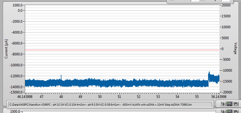
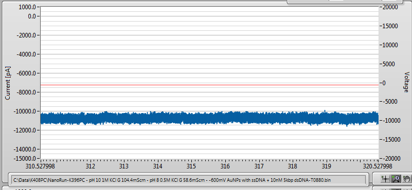
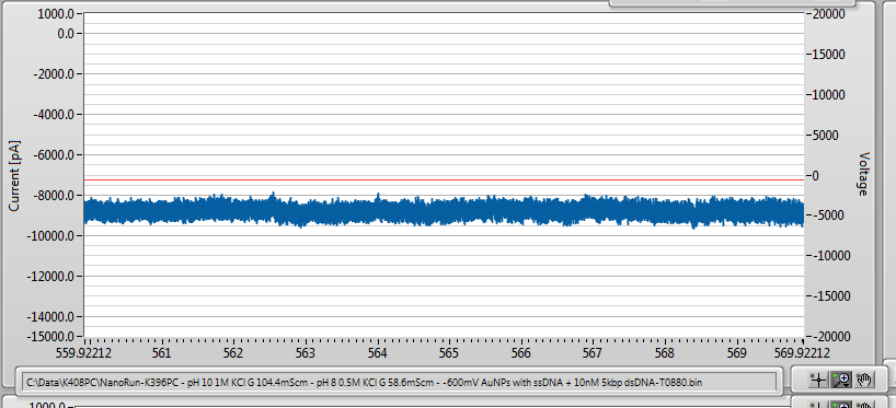
The pores I tried to use were from 10-20 nm in size. In hindsight, now that we have characterized the beads better, this is perhaps not too surprising. Even though the charge on the beads is huge, the high DNA density means that the effective size is such that the DNA strands cannot penetrate very far into the pore at all. Since the electric field is heavily localized within the nanopore, the actual force felt by the beads is probably not very big. Assuming a rigid sphere of 75-nm diameter, it would only penetrate ~3-nm into the 20-nm pore, and would only sample the edge of the electric field. With this in mind, it is possible that the baseline fluctations I consistently see in the regular chip experiments really are approaches by gold nanoparticles, blocking some of the access region without actually really clogging the pore.
It seems likely that using smaller gold particles will be better, for a number of reasons. First, the smaller particles are more stable in suspension, and will allow us to push the salt concentrations higher. The smaller diameter means larger curvature, which will promote insertion of the attached DNA into the pore, giving a stronger force to clog the pore. Once we have finished off the current set of 50-nm particles (we have 8 or so kits left, each good for ~2 experiments) I will purchase some 30-nm gold particles instead. This will give something like 50-nm effective diameter with ssDNA conjugated, which should still be excluded by the filter.
Since the regular chip/gold particle experiments weren’t working out, I switched back to the NPN chips to try to get some controls with ssDNA. Here, I actually get some pretty cool results. In this experiment, I have a chip with a filter, as shown below.
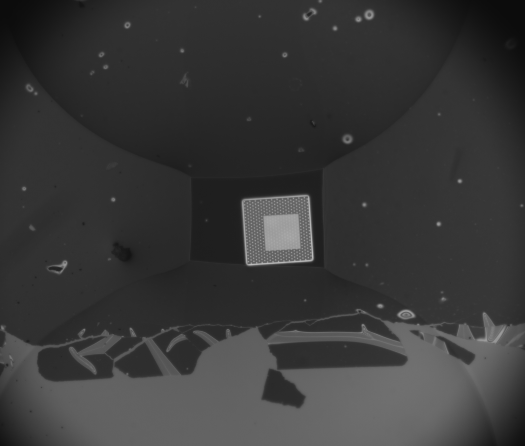
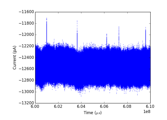
The chip is plasma cleaned to make it hydrophilic and to bond it to silicone gaskets. It is chilled to promote condensation, then wetted using the vaporizer. At the start of the experiment the pore is 11-nm, but it is unstable as soon as I add KCl and grows during the experiment to about 30-nm over several hours. Nonetheless, the noise performance is excellent:
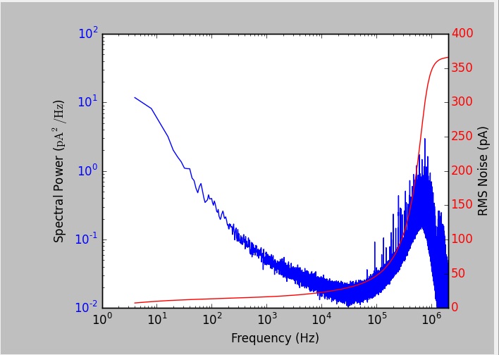
I am using the Chimera VC100 for by data acquisition now, which allows me to get 1MHz bandwidth instead of the usual 100kHz from before. For actually analyzing data I end up filtering down to a few hundred kHz, depending on the signals I am seeing.
First experiment was to add 180nM 50-nt thiolated but non-reduced ssDNA. We immediately see signals through the pore.So, we finally have a positive experiment showing diffusion of short DNA through the filter!
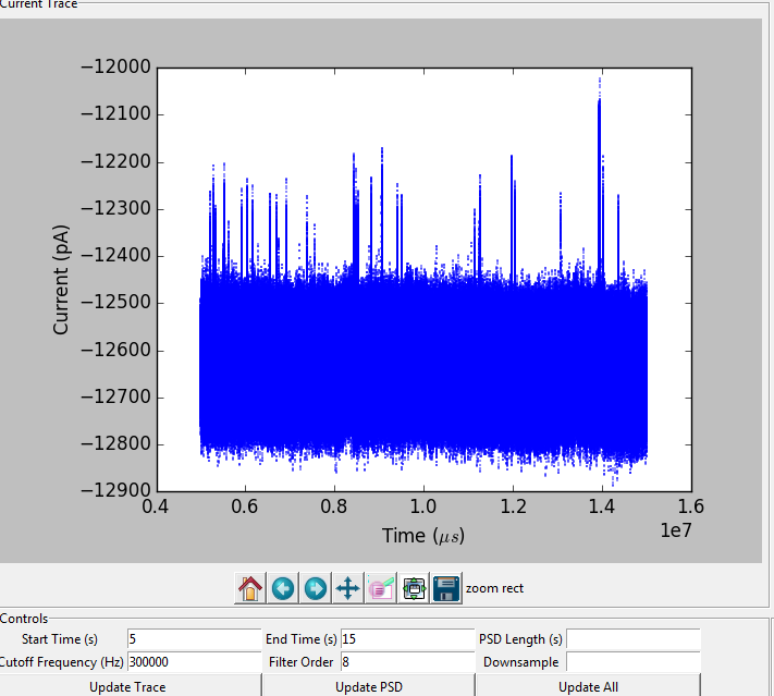
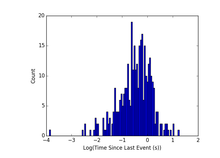
It gets better, though. Over the course of the experiment, the event rate increased dramatically with time. It started out around 1-10 events per second (histogram of time between events for the first ~200s of data below) which is about par for 180nM concentration using the axopatch.
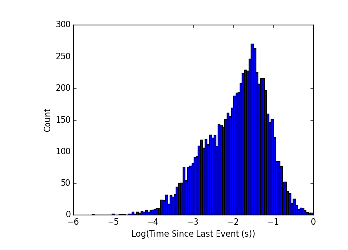
By the end of the experiment, however, the events rate had increased by a factor of 10 or more (last 200s of the experiment):
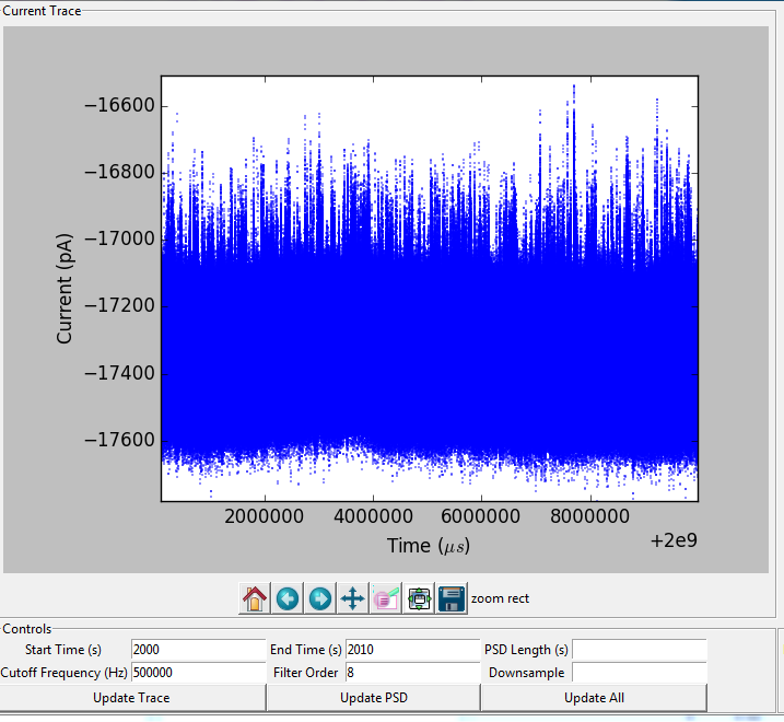
The events at the end are so dense it’s hard to see them on this scale anymore.
I think what we are seeing is the effect of diffusion through the NPN. I pre-mix my samples before injecting them, so the concentration is homogeneous on the outside of the NPN layer. The capture rate is linear in concentration near the pore, so the increased capture rate should correlate well with the rate of diffusion through the membrane. We can probably knock together a simple model and compare numbers in the near future. Unfortunately, this pore grew quite a bit over the course of the experiment. Capture rate should be related to the pore size, so that might account for some of this, meaning that we probably can’t get a good diffusion rate from this experiment. But it’s something to bear in mind going forward for when I get a stable experiment going.
Next, I added a mix of gold nanoparticles and 180nM ssDNA. At this point the pore was so large that I could only use 80mV on the Chimera without saturating the system, so it’s not quite directly comparable to the first experiment. Sadly, I did see some clogging.
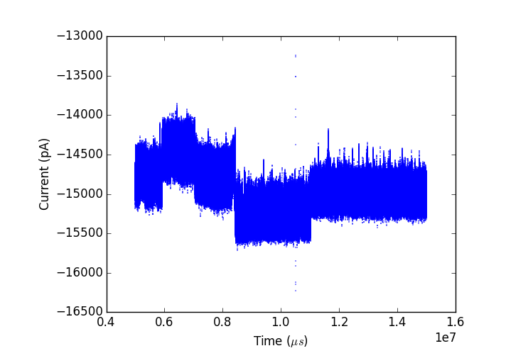
However, the pore had been fluctuating pretty wildly as it grew, so I am going to call this inconclusive on blocking nanoparticles and wait to reproduce it before making any claims one way or the other.
After the experiment was done, I tried to dismount and image it again to verify the integrity of the filter layer. Several things went wrong at this point. The PDMS layer was bonded more strongly to the gasket than the chip, and peeled off with the gasket. You can see the gasket/PDMS layer along with most of the NPN below (imaged from the bottom side, previously attached to the chip).
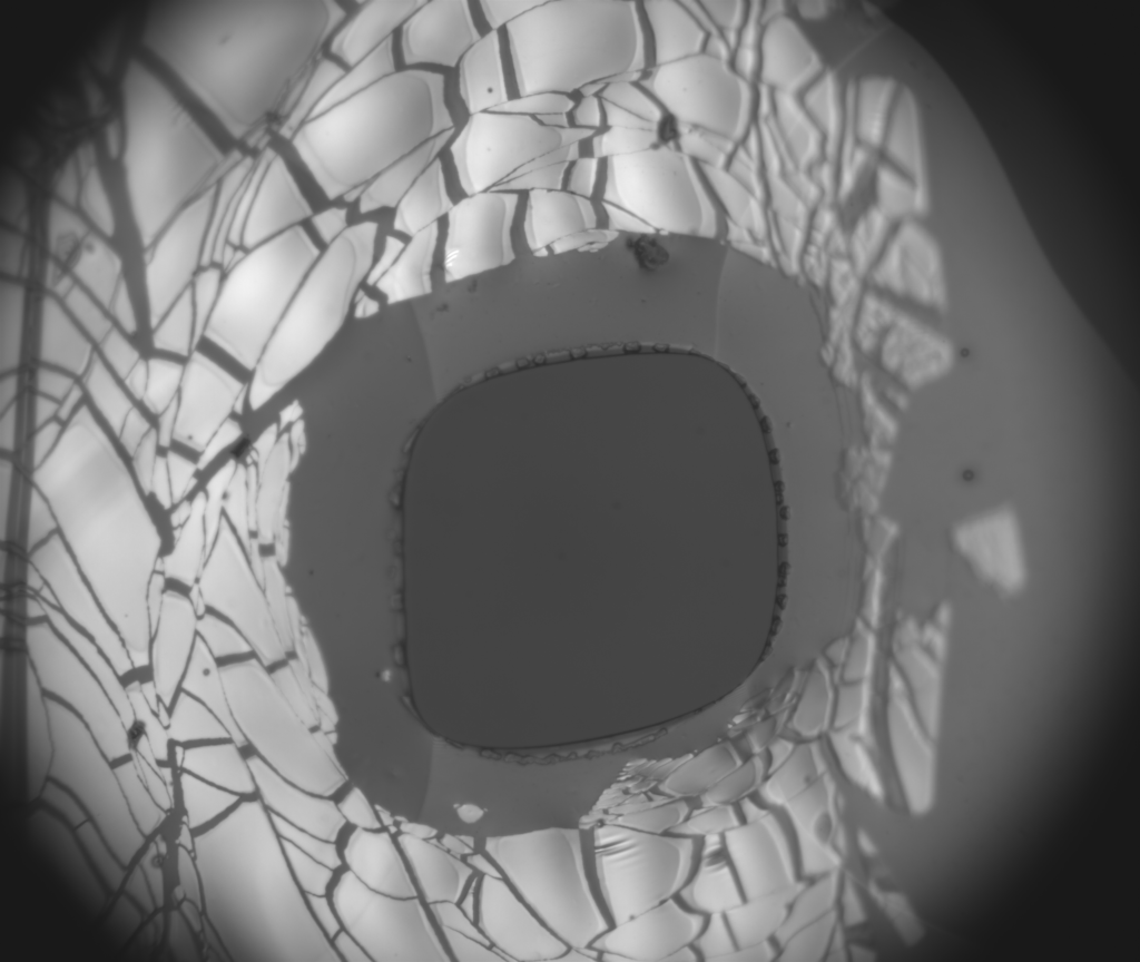
The chip cracked into several pieces as I got it out of the flow cell. This batch of chips is slightly larger than the previous one, and just barely fits in my flow cell, so the tight fit making snapping the chips a big risk.
However, despite all this, I was still able to verify the NPN filter layer. Because the PDMS did not peel off the NPN over the membrane, it was still visible, and shows no wrinkles or liftoff:
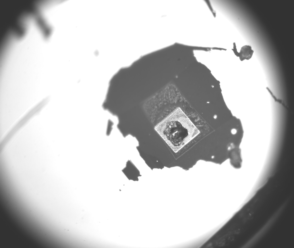
The membrane was destroyed with the chip cracked, but the NPN layer still covers it completely, so I am confident in saying that we just saw DNA going through the filter layer.
I also ran a control experiment without the filter. This pore also grew a bit during the experiment, but like the other one showed excellent noise performance. With 180nM ssDNA, I get signals like this:
The event rate is pretty low, but it stays consistent over the whole 2000-s experiment. This supports the idea that the increase in capture rate in the filter case was due to diffusion through the filter. This kind of pore-to-pore variability in capture rate is not uncommon.
Once I added gold nanoparticles to the system, I got clogging. For this I went back to the axopatch since I don’t need high bandwidth to resolve long clogs.
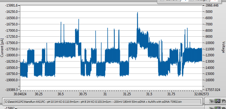
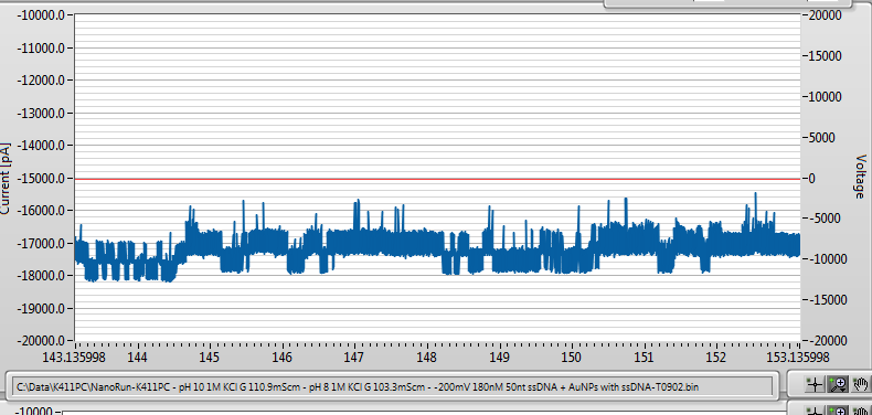
The pore eventually clogged permanently (ie, entered a lower conductance state). Interestingly, the clogged state still showed passage of ssDNA, indicating that the pore was still accessible to DNA, consistent with a gold nanoparticle being in the access region and partially blocking the opening, without actually fully inserting and clogging. One potentially confounding fact is that the step changes we see in the conductance are consistent with blockages from ssDNA, so it’s hard to be sure this is gold nanoparticles doing the clogging rather than just several stuck ssDNA strands.
Very promising results overall. Still needs to be reproduced several times, and there is still a fair bit of work to do characterizing gold nanoparticles and optimizing their size and the salt conditions to be used. For the next round of experiments, with and without a filter, I am going to add gold nanoparticles first. When ssDNA has been running for a long time in the pore, clogs get progressively more likely, so I need to rule out the possibility that the clogs we see are ssDNA alone by running the AuNPs first.
Questions/Comments/Thoughts/Suggestions?
Outstanding!
How might NPN improve capture rates? A filter should hinder diffusion not help it, although with NPN we expect the hindrance to be minimal. Perhaps NPN is selecting ssDNA that is able to move to pores more quickly or enter more smoothly?
Perhaps it is unrealistic to think we can eliminate clogging with NPN at this stage. Especially since either the particles or ssDNA can do it. There is an optimization effort in our future where we adjust NPN pore sizes and gap height. Right now we hope to show we can prolong the lifetime of pores compared to no-filter. Can you say anything about that from these experiments?
Also what happened to the problem of membrane fluctuation above the oxdide spacer? Was this the material with the SiN cap or does it just not matter when things are rolling like this?
I don’t think NPN is improving the capture rate – different pores have different capture rates for reasons that are poorly understood, I think it just happened to be a good one with NPN and a (relatively) bad one without it.
This was the material with the nitride cap. I saw a small amount of baseline fluctuation before the experiment, but during the experiment the pore was growing steadily so it’s hard to distinguish fluctuation from growth.
I don’t think I can say much about lifetimes from one experiment – this one probably could have gone longer if the pore size was stable. It’s a nice proof of principle, but in many ways it was not an ideal experiment.
Nice Work! It’s exciting to see DNA make it through the filter. You mention in the post that the pore widens with time as the experiment runs (from the E-field causing additional breakdown?). Is it possible that the NPN tent is experiencing some of the same widening? If it is, perhaps we see increased events because there are more holes to make it through the tent. It seems like a catch-22; we want the tent to be close enough to be within the capture radius of the pore, but be stable enough to give us good screening characteristics. Would a nanoporous tent made of a different material (non-silicon) be useful to you?
The pore widening is not the result of the electric field (200mV that I used for the DNA sensing experiment is far too small to cause breakdown in reasonable timescales). The pore widening and instability seems to be a joint property of the membrane material and the solution, since I generally find less instability in LiCl than KCl, and some nanopores are more stable than others even in the same solution. Note that this is not unique to the filter chips, all nanopores have this problem to some extent, it’s all just a matter of timescale. THe voltage drop across the NPN is so small that it is definitely not affecting the NPN pore size – but I can’t rule out natural instability of the pores in solution.
The voltage drop across the NPN is so small compared to the voltage drop across the single pore that being in the capture radius is maybe less important than I thought originally (hard to say without more experiments). Bottom line is that we seem to be relying pretty heavily on diffusion to get DNA through the filter rather than actively pulling it through. Doing experiments with DNA length between 50-1000bp might give us a better idea what’s going on, but that’s an experiment for later.
A tent made of different material could potentially be useful if we think that NPN stability is an issue. Maybe if I cleaned off a used filter and sent it back to you for imaging we could check if the pore size distribution shifts significantly over the course of my experiments?