Preliminary Data on Gold Nanoparticle Separations
Using wafer 302 I set up 2 20nm and 2 50nm gold diffusion experiments. After 24 hrs I removed the retentate and filtrate and looked for the gold using the Malvern Zetasizer. We can’t perform the diffusion with both 20nm and 50nm gold because the Zetasizer cannot differentiate between the two sizes.
|
r value |
Sample |
Gold in Retentate? |
Gold in Filtrate? |
|
3.6 |
20 nm |
Yes |
No |
|
3.2 |
20 nm |
No* |
No |
|
3.6 |
50 nm |
Yes |
No |
|
4.5 |
50 nm |
broken |
broken |
*It was difficult to collect a big enough drop from the retentate of the second sample, and the gold may have been diluted beyond the Zetasizer sensitivity.
I went back to test the Zetasizer sensitivity, and it appears that if less than 10% of the starting volume diffuses across the membrane, we won’t be able to see it (this means a 1:300 dilution or more of the starting solution).
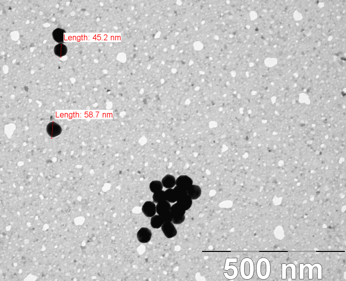
I had a couple of TEM pictures taken of my gold samples on wafer 214. The pores sizes on this wafer are similar to 302, and you can see while 20 nm appears to be small enough to fit through the pores, 50 nm does not.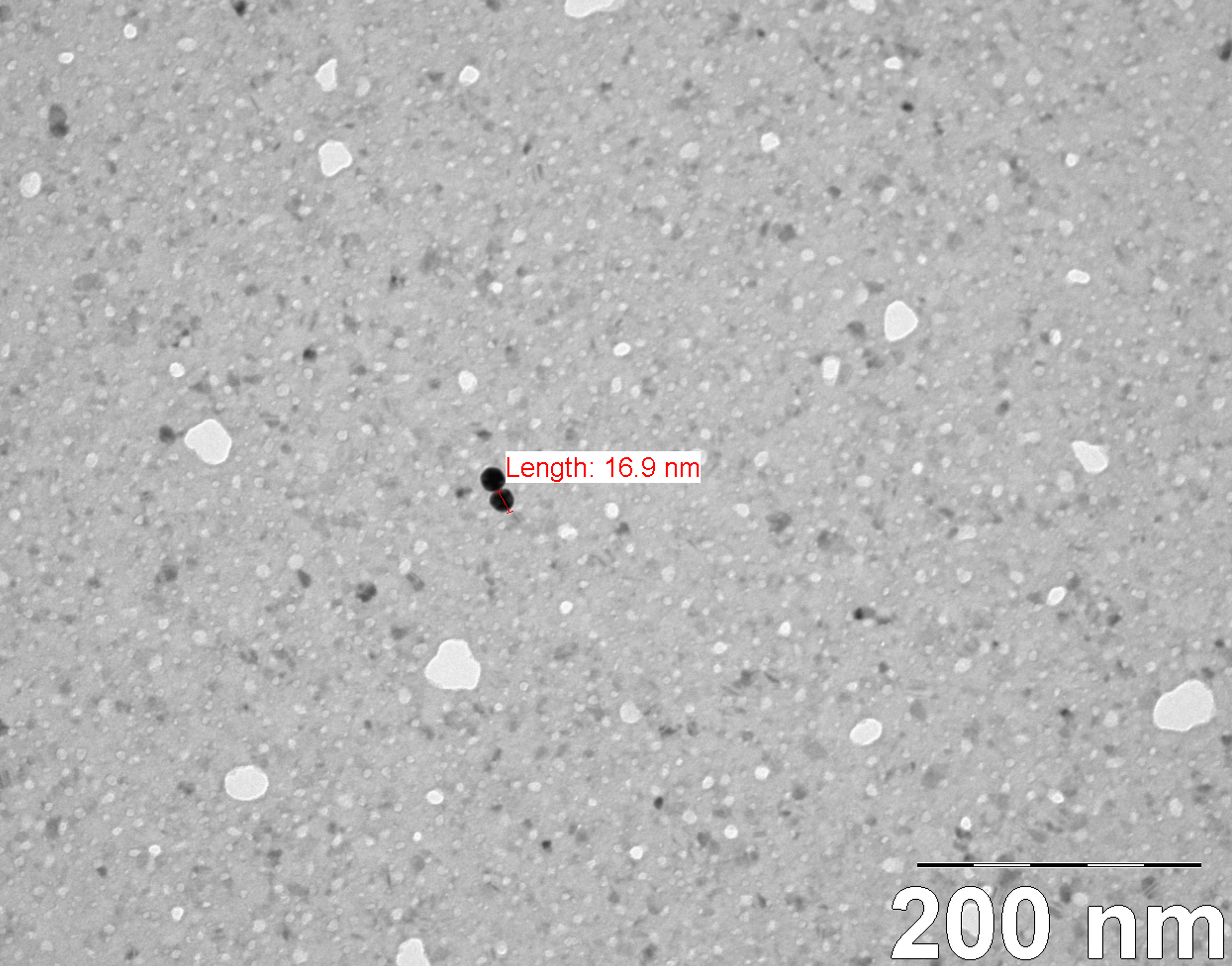
So why doesn’t 20 nm gold pass through the membrane in 24 hours? Is it passing but we just can’t detect it yet? Is there a better way to detect passage? Tom suggested using quantum dots and detecting the composition of retentate and filtrate with fluorescence. Next up I’ll be testing our 10 nm and 5 nm gold for diffusion using the Zetasizer.
electrostatics? are the Au particles in DI or a salt solution?
Why can’t the Zetasizer distinguish 20nm from 50nm gold. These are much larger than proteins, have a high scattering/extinction coefficient, and differ by more than 2X in diameter (>8X molecular weight)? This would seem to be a no-brainer for this tool, but I really have no idea how it exactly works….
I agree that electrostatics would be an issue (what is the molarity of the buffer?), and rigid spheres may also behave differently in our pores. I would also guess that the diffusion coefficient would be smaller than proteins, as these NPs are quite a bit larger and more dense? It seems like the physics of the random walk of a solid metal sphere would be much different than a soft, folded protein, but maybe not. Also, in your posted TEM images, it’s interesting that they tend to be paired up or clumped. Perhaps they are starting to clump in solution (and this would apparently be invisible to the Zetasizer)?
Is there a reason why you cannot just run all the NP sizes at the same time in parallel overnight experiments, with several samples of each? I have no feeling for how complex these experiments are? Something has to go through eventually.
Yes electrostatics is an issue here. Gold particles clump together in high salts, so I’ve been diffusing them into 20mM HEPES (PBS is about 150mM salts). The initial solution is in DI H20. I imagine shielding is an issue.
The Zetasizer has an inability to distinguish between two particles if they are too close in size (I think the original salesman said they needed to be something like a magnitude different). Larger objects scatter to such a higher degree that it’s difficult for the software to recognize and calculate the distribution of a smaller species if there’s any overlap in the intensities.
If the particles clump it is immediately apparent using the Zetasizer. The solutions I used for the TEM samples were a bit too dilute, and the observed clumps were easier to pick out. I did find some singular 20nm particles. The clumping observed on the TEM could be a result of drying also.
I can run up to 4 samples at once using my setup. This experiment was 2 20nm and 2 50nm, although one of the 50s broke. I plan on doing 2 10nm and 2 5nm next. Doing one of each in parallel means there’s a higher chance of losing one of those samples due to breakage. The next 4 tests will be on the same wafer with the same conditions, so should be comparable. I can’t do time points with this experiment because I need the entire sample (60ul) to test with the Zetasizer.