Endothelial cell attachment to porous substrates via focal adhesions is initially affected by pore spacing and not absolute porosity. Maturation of endothelial monolayers via cell-cell interactions occurred over the same time course regardless of substrate porosity, pore size and contact area. Cell-matrix interactions at maturity were dictated by substrate stiffness.
Data
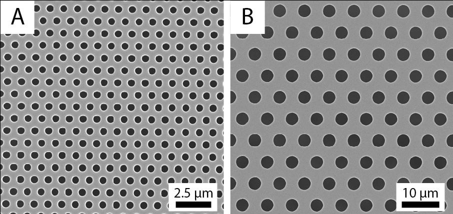
Fig. 1 SEM images of 0.5 mm and 3.0 mm pore size membranes. Spacing between pores is one pore diameter. Porosity for both membranes is 22.7%.
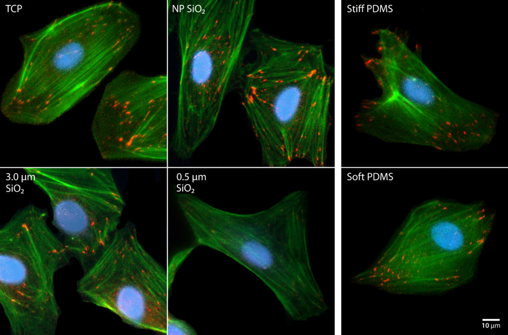
Fig. 2 Cell attachment to substrates through focal adhesions at 24 hours. HUVEC were cultured on tissue culture plastic (TCP), non-porous, 3.0 µm high porosity, 0.5 µm high porosity SiO2 membranes, and stiff (E = 2 MPa), and soft PDMS (E = 5kPa). Cells were stained with DAPI (nuclei, blue), phalloidin (actin filaments, green), and anti-vinculin (focal adhesion vinculin, red).
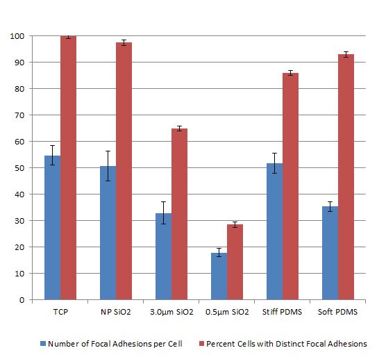
Fig. 3 Quantification of focal adhesion formation. Formation of distinct focal adhesions was quantified for all substrates – number of focal adhesions per cell and percent of cells with distinct focal adhesions. Error bars are +/- SD. Sample size >50 for each substrate.
[missing figure]
Fig. 4 Endothelial Barrier Maturity. HUVEC were cultured to form a monolayer on tissue culture plastic (TCP), non-porous, 3.0 µm high porosity, 0.5 µm high porosity SiO2 membranes, and stiff (E = 2 MPa), and soft PDMS (E = 5kPa). Cells were stained with DAPI (nuclei, blue), anti-VE-cadherin (cell-cell junctions, green), and anti-vinculin (focal adhesions, red) to visualize formation of endothelial barriers.
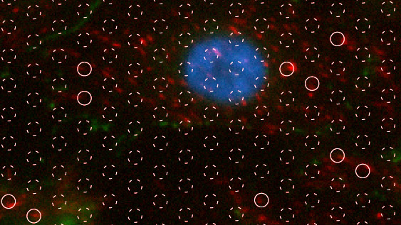
Fig. 5 High-resolution fluorescence image of focal adhesion formation on 3.0 µm pore size SiO2 membranes upon reaching confluence. White circles are locations of pores. Solid circles are locations where focal adhesions (vinculin, red) are present over the pore or at the pore edge.
Supplemental Data
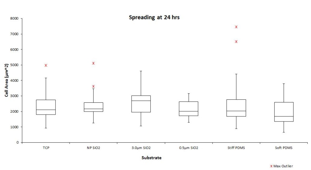
Fig. S1 Cell Spread Area at 24 hrs. HUVEC spread area was quantified 24 hrs post-seeding on TCP, non-porous SiO2 membranes, 3.0 µm pore size SiO2, 0.5 µm pore size SiO2, stiff PDMS (E = 2 MPa), and soft PDMS (E = 5kPa). The box plots represent median and IQR of spread area. Whiskers are +/- 1.5IQR.
In this post, I would like to sum up all different annealing scenarios, which corresponding to different morphologies of the pnc-Si membrane so far we have observed. It should be very helpful to understand the evolution of the pore formation during the annealing. This whole evolution I believe, not only applies to the oxide/silicon/oxide stack,…
Submitted on April 7, 2014. Posted to NRG Cloud under NRG/Funding. Figure 2. Dead End Filtration with SEPCONTM (A) SiMPore’s SEPCONTM spin cup with a silicon nanomembrane chip. B) Dead end filtration in a centrifuge. The direction of the centrifugal force is indicated with the gravity vector g. Figure 3. Nanoporous Silicon Nitride (A) pnc-Si…
Over the past week Andrea and I designed a new co-culture support structure for use with SiMPore’s 5.5 mm square membranes. The devices are built for use in standard 24-well plates and should prove useful to anyone wanting to do basic co-culture with these membranes going forward. The device is constructed of PDMS rings as…
As per Chris request, here is a photo with a brief description. Sorry the image is still not good… the surface of the plastic reflects lights and it is transparent, plus I am using a poor camera. But I hope the description is useful and anyway tomorrow we can check it out in the meeting!
I am doing a few more membrane transfers and trying to troubleshoot my delamination problems, and I wanted to run my protocol by you guys before I mess up any more chips. I was able to get about 3/7 transfers visibly very good without any wrinkles, but they all still delaminate, either during the fabrication…
Anticipating the eventual introduction of nanoporous SiO2 can we call non-porous SiO2 something other than np-SiO2?