To follow up on my recent posts about HUVEC YAP localization after 24 and 72 hours (https://trace-bmps.org/blog/data/2016/10/19/yaptaz-passage-one-staining and https://trace-bmps.org/blog/data/2016/10/11/yaptaz-staining-on-membranes-and-nonporous-sio2) , we conducted the same study using Adipose Derived Stem Cells. Due to the fact that these are mesenchymal stem cells, we thought that their expression of YAP might be a bit different due to the fact that HUVEC YAP and ADSC YAP seem to have slightly different functions. To reiterate on the basic information about YAP, it’s related to a mechanosensing pathway and the YAP localizes in the nucleus when the cells “sense” a stiff substrate and it localizes in the cytoplasm when they “sense” a soft substrate. In the HUVEC studies, we saw that the nuclear:cytoplasmic YAP decreased on porous substrates but none of the porous substrates were significantly different from each other, while all were different from nonporous SiO2 and TCPS. To gather this information with ADSCs I did the following:
Cyto-Vu devices were made using the above substrates and 300 ADSCs were seeded per membrane. The cells were allowed to adhere to the substrate for 1 hour and then the wells were flooded with 800uL of media. After 24 hours or 3 days, the cells were fixed in 3.7% formaldehyde for 15 minutes, permeabilized with 0.1% Triton T-100 for 3 minutes, washed with 4% BSA for 15 minutes, and 50 μL of the YAP antibody (0.2mg/mL was diluted 1:100 in PBS) was added ontop of the substrate for half an hour.
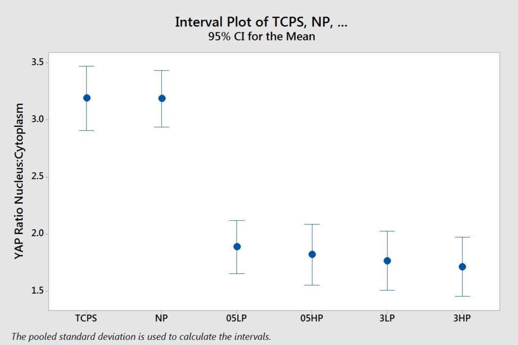
The images were acquired and analyzed in image j. The nucleus’ brightness was measured as well as the background and the cytoplasm of the cell. To analyze the data, the background was subtracted from the values for the nucleus and the cytoplasm. Then, the intensity of the nucleus was divided by the intensity of the cytoplasm. Sample sizes were at least 20 cells. The results are as follows:
24 Hours
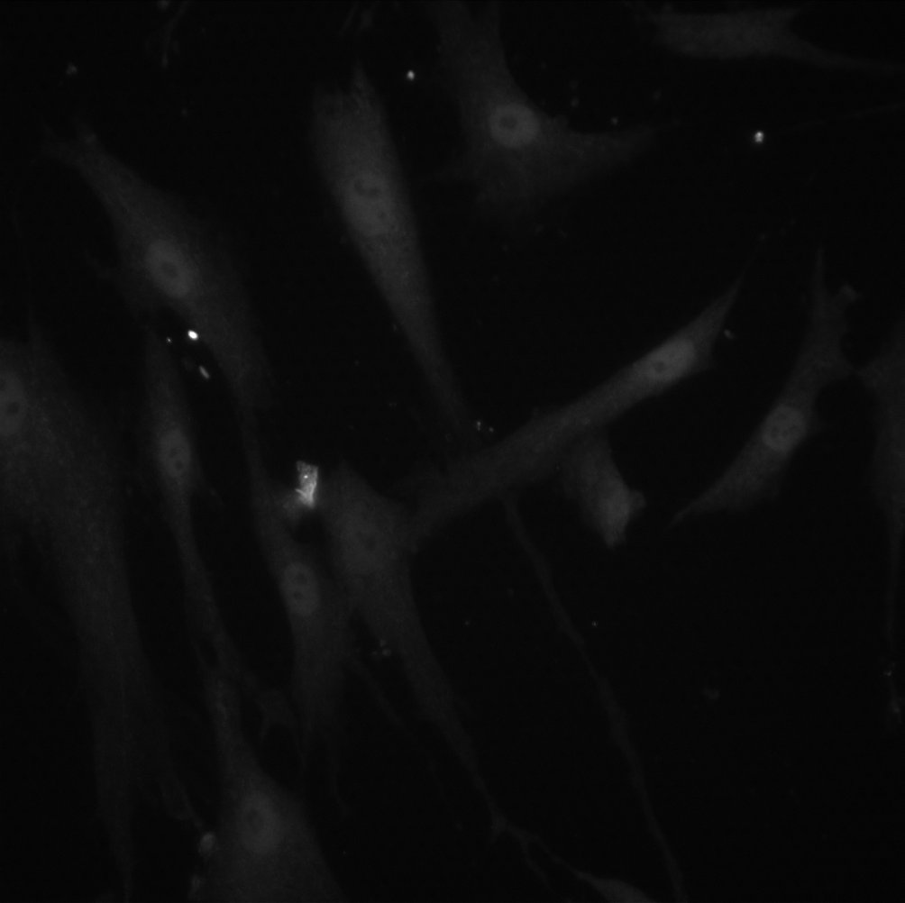
TCPS: 3μm HP:
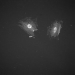
Both images are at 40x
72 Hours
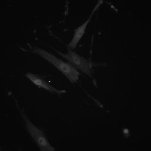
TCPS: 3μm HP:
Both images are at 40x
The take home from this data is that the ADSCs exhibit a similar trend as the HUVECs do in regards to YAP expression over 24 and 72 hours. The two porous substrates are not statistically different from each other and the porous substrates are not statistically different from each other. However, all of the porous substrates are statistically different (lower) than the nonporous substrates. This follows with our previous data in insinuating that the membrane discontinuous surface area may be impacting the cells similarly to soft substrates. It is interesting to note how pore size and porosity did not impact the YAP expression in any statistically relevant way. Up next is ADSC memory experiments where we will compare this data to ADSCs that have been on TCP for 10 days (the longest time point in the study mentioned in a previous post). Literature would suggest that YAP expression would be the same on all substrates if this “memory” phenomenon was true but it will be interesting to see if “memory” is exhibited on our membranes.
Thanks for reading!