Membrane Discoloration
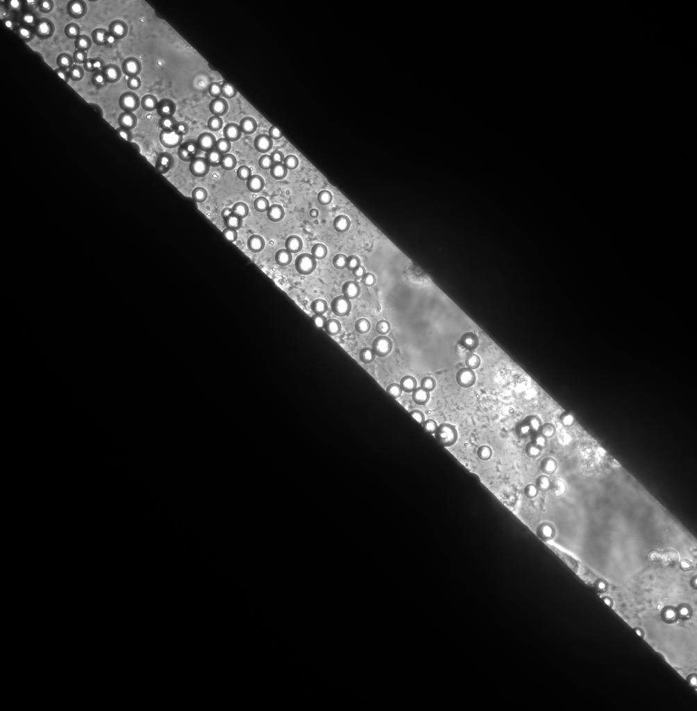 15um beads on discolored membrane surface, having some slits where the beads are not found
 The localization of microbeads suggest that they have fallen across the slits in the membrane
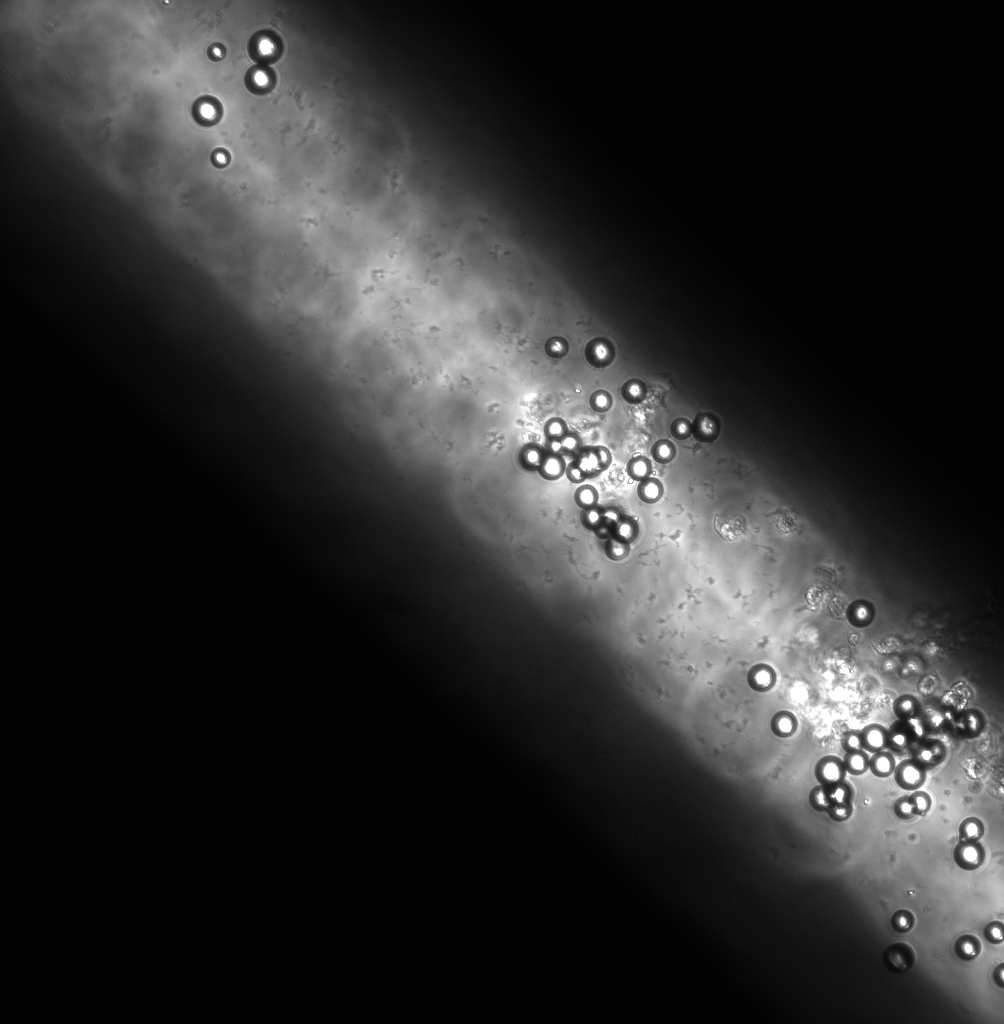
 Transparent membrane with microbeads on its surface
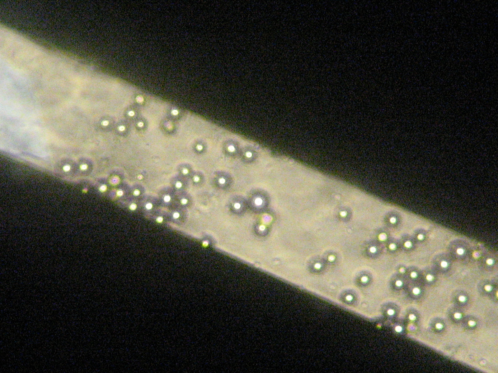
 These membranes are not treated with cell media, hence are not discolored. There’s a slit which appears to be same as the transparent membrane (below)
 Above shows the image of the beads that have crossed the broken membrane and lie on the other side
Now its confirmed that the membranes are discoloring and getting converted to something like glass. I carried out some experiments using 15um microbeads and the experiments suggested that the membranes were intact but looked as if they were not present, i.e they were transparent! Till now we’ve been thinking that the membranes are degrading in the cell growth media, which might not be the case. Instead, the membranes just went transparent. I dropped some microbeads on the media treated membranes, which I thought to have disrupted, but observed that the beads stayed on the membrane height. There might have been some slits on the membrane due to which the beads crossed and fell on the other side. The localization of the beads on the other side confirmed that the membranes were present and their transparency mislead to think that they were gone. The non media treated membranes have a yellowish tinge as shown in one of the figures and this is how I used to determine whether the membranes were intact. But the membranes lose this yellowish tinge and turn transparent. The appearance of discolored membrane is the same as that of a broken membrane which I used as a control. and found that the beads pass easily across the broken membrane.
Further, there’s a need to find an alternative to determine the presence of these transparent membranes as everytime this microbead test cannot be carried out. Also, this might have resulted due to the UV ozone treatment of the membranes.
I don’t completely understand your images (what is the one with the blurry membrane?), but I think you are correct that the membranes are oxidizing, not dissolving. It’s nice to have proof.
The last image looks like the beads are still on a membrane – is this one mixed up with the one showing the blurry membrane. Perhaps you can explain at the next meeting.
Even these membranes may be visible if you image in reflection at a wavelength where the water and glass have the most significant refractive index difference. It will be tough, though….
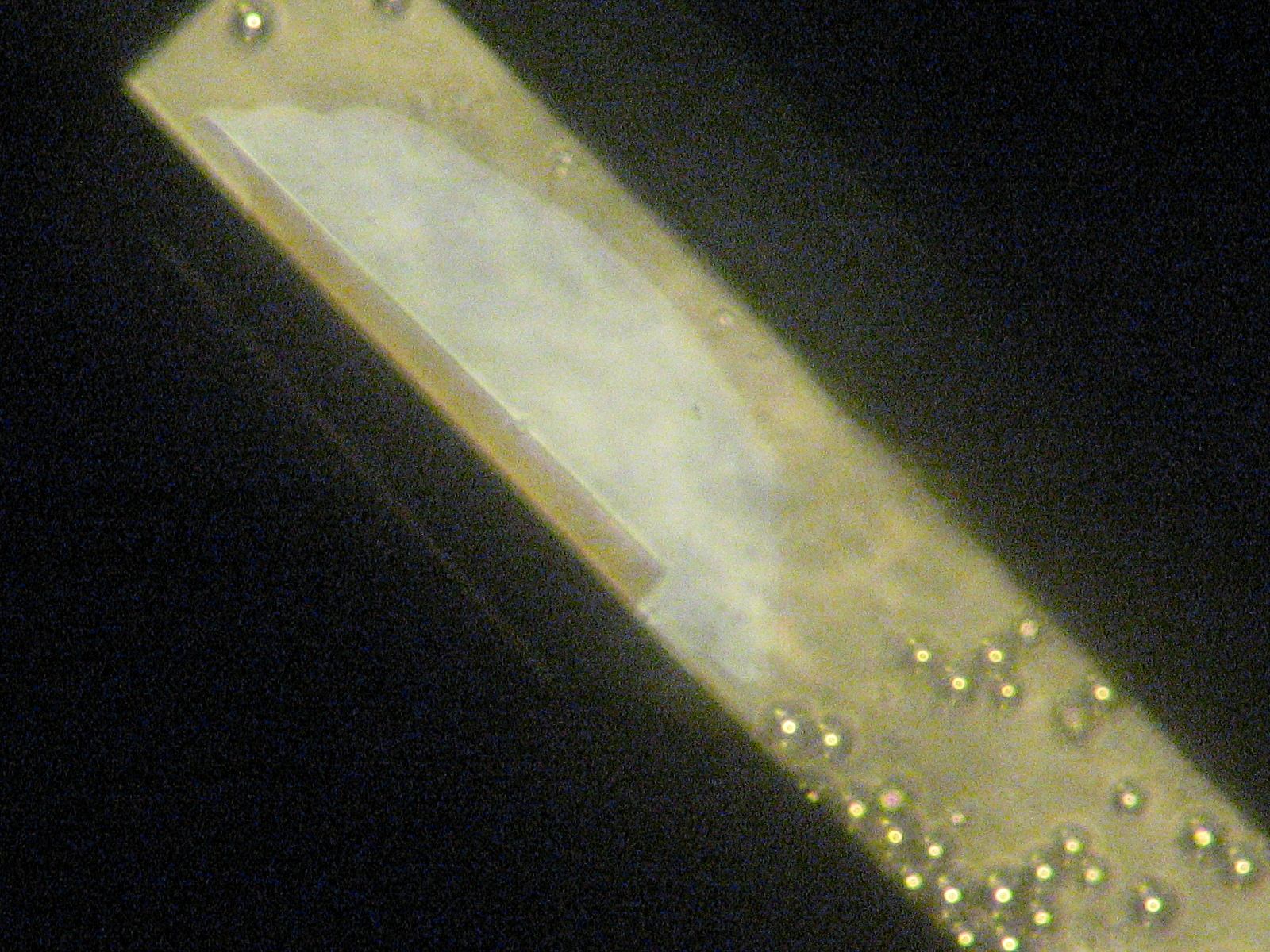
The blurry membrane image (figure 2)shows the bottom of the culture plate in which the membrane was kept. Since the membrane is broken, the beads pass through and settle at bottom. The membrane slit is out of focus to show the beads at the bottom. I hope this makes things clear or else I’ll try explaining it in the meeting. You’re right, the last image looks mixed up, I guess I picked up the wrong image in place. But still it doesn’t really show the beads on membrane surface. anyway…