Microscopy on TEM Grids with Kevin Webb @ University of Nottingham
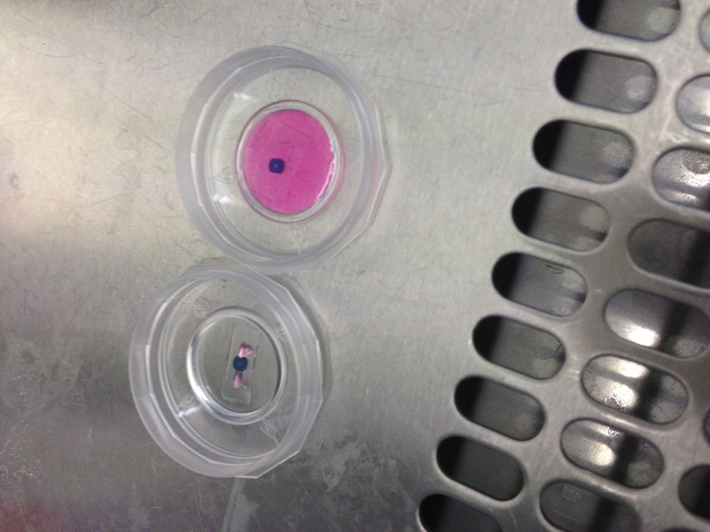
Dr. Webb is very interested in the thin nanomembranes that our group experiments with. To date, he has been trying to get cells to grow on SiN microporous material and image them with a few different types of microscopy (Phase, Confocal, Scanning Conductance Ion Microscopy). The cells have not grown well natively on the nitride membranes, or with simple collagen. To try and get some cells to grow, we have used a 500×500 micron TEM (pnc-Si) to culture cells in an open device. Rat neural progenitor cells were incubated for 16hrs and then observed under a phase/fluorescence microscope for ~30 min, when it became apparent that the cells were slowly balling up and becoming unhappier. The cells were then stained and fixed. Phase images were obtained, and overlaid with a fluorescence signal from the stain (correlating to the nuclei of the cells). The cells were then imaged using confocal microscopy (DIC contrast), using a z-stack with a slice volume of 4 microns. Finally, the cells were imaged using SCIM.
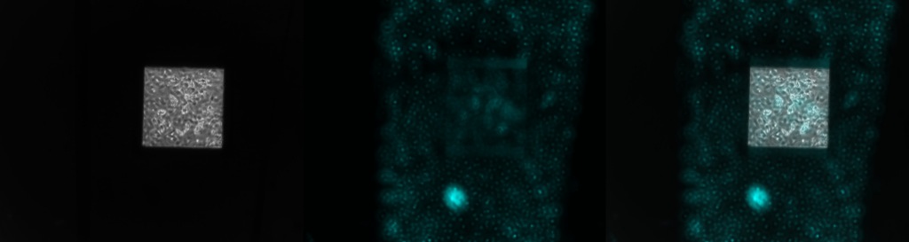
It’s possible to capture the entire window with a 10x objective. This makes identifying particular cells within the window entirely feasible for specific discrimination and follow-up imaging.


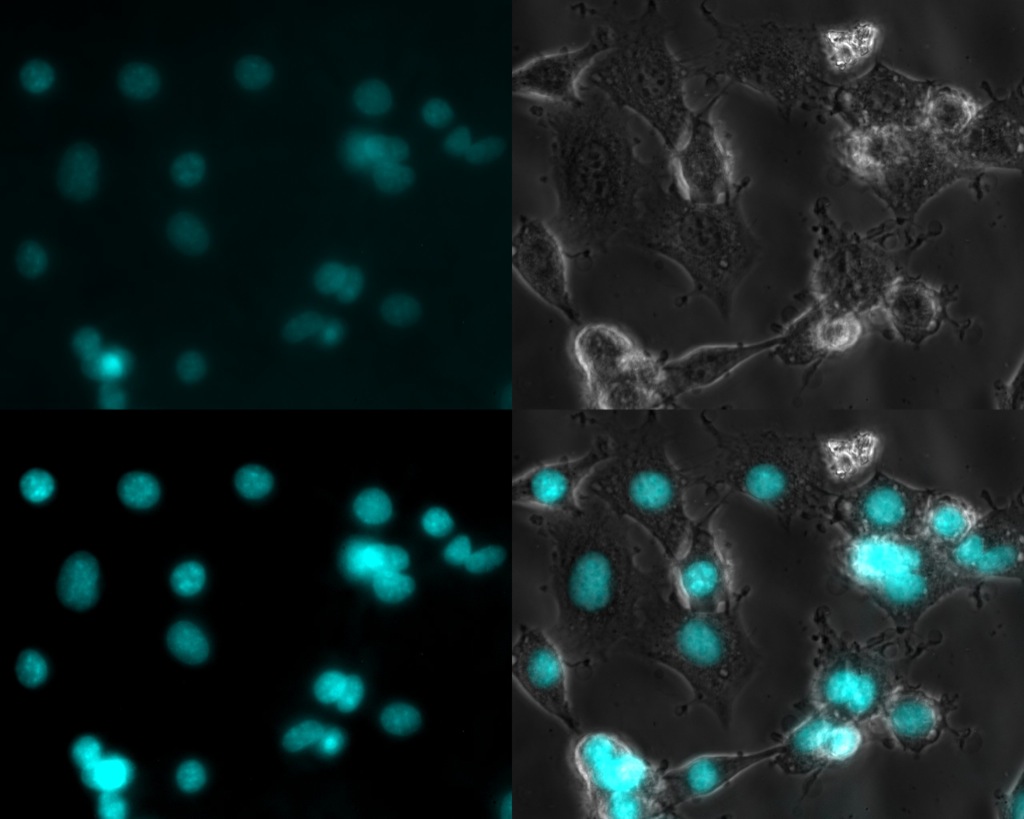
The cells existing in the bottom of the membrane were very nice and happy. Some dendritic growth and outreach is very obvious, very similar across the whole channel.
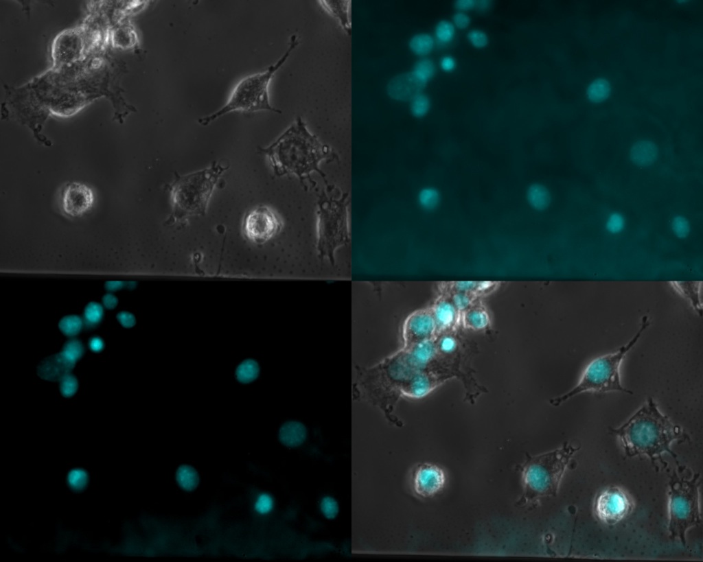
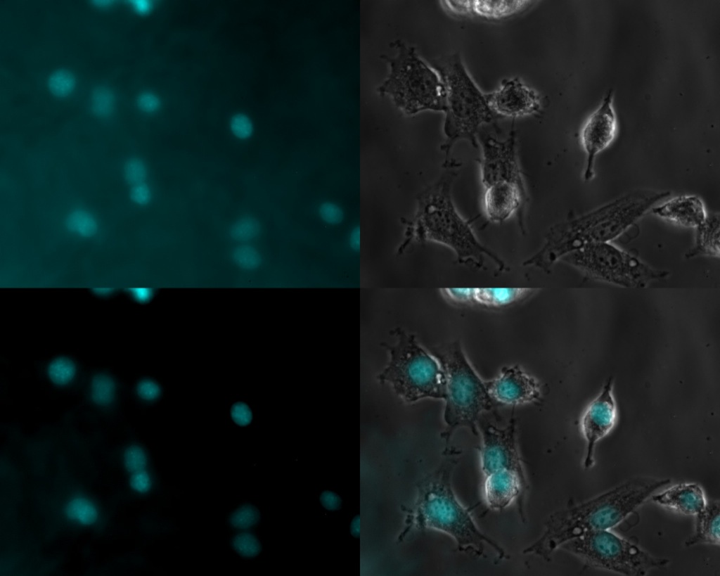
Imaging the cells through the filter on the flat side of the TEM grid was difficult. The UV fluorescence signal took a double hit (illuminating and detecting), resulting in poor contrast. It took about 10x intensity compared to the bottom channel to get these images. Cleaning up the signal produced a nice overlay,but it is far from optimal imaging. TEOS nanomembranes would have a significant impact on lessening the impact of the pnc-Si UV absorption. These membranes have not been post-annealed, so the only boundaries are the native oxide-si-native oxide.
We created a z-stack using DIC-confocal imaging to image the cells on the flat of the membrane and in the channel below. We made a 107 images of ~4 micron thickness, corresponding to the 400 microns of separation between the membrane and the bottom of the channel. I assembled these images in ImageJ to create the reconstructions you see here.
Our initial attempts to work on the SCIM did not go smoothly. The principle operation of the SCIM is much like an AFM. In tapping mode, the probe oscillates at a known frequency and speed. When the tip comes close to the surface of a cell/object, the hole within the tip cannot get the same access to electrolyte as when it is further away. Therefore, the electrical resistance of the pathway is increased. Feedback keeps the tip from dragging along the surface and ruining the tip. In our initial attempts, the feedback did not prevent the tip from running through the membrane and tearing it. It is possible that the membrane has sufficient permeance to prevent the resistance increasing when the tip proceeds to the surface. As I left overnight, a slower scan may have produced some useful results without tearing the membrane, but they aren’t ready to show yet.
Ultimately, the cells on the membrane did not look as happy as cells on the glass. Kevin will be trying different coatings to make the cells adhere better. Fibronectin worked well for my endothelial cells, collagen and poly-l-lysine are also other candidates.
I’ve never really understood the various surface treatments of our membranes for cell growth. How do you characterize how much of these treatments actually sticks to the surface and how do you optimize it? If treatments don’t work, it’s probably because very little adsorbed to the surface. SiN is not very reactive at all, so it’s no surprise that very little material adsorbs to the surface.
With appropriate surface functionalization, you should should be able to convert SiN into whatever you want and we have the tools to do this. IT seems like it would be more efficient to use directed attachment to optimize these surfaces, as needed…
Interesting work so far…Thanks!
Hi all, great to see the images up and to get online with the group 🙂
Thanks to Jim for the access, and to Greg for the nice writeup. SICM images and more to follow soon.
Chris – if you have some ideas on how to functionalise these surfaces so that biomolecules can adhere better (collagen, fibronectin etc) we’d love to hear them! In Nottingham I have access to a TOF-SIMS (time of flight secondary ion mass spectrometer) which we could in principle use to quantify bound molecules, and potentially correlate this with contact-angle measurements. What do you think?
All the best
Kevin – we have various vapor-phase methods to silanize silicon-based surfaces. For example, we can silanize the surface and leave amine terminations, or various other leaving groups, provided silanes with the right characteristics can be found. Alternatively, we can leave the surfaces amine-reactive, so that proteins in a solution will covalently bind to the surface. Chemically reactive surfaces tend to loose activity quickly, though, so subsequent treatment needs to be done within a few hours, and then potentially stabilized depending on the robustness of the molecules.
Once the various form-factor and optical performance issues are worked out, we can potentially try a couple surface chemistries to improve cell adhesion and growth. I’ve never done TOF-SIMS, but if it can be quantitative over a small membrane, it could be useful. Thank you.